
Page 76 - 2018_09-Mondo
P. 76
D. Wu et al.
PARP) are increased after isobavachalcone treatment. Given that DHODH represented the rate-limiting step of de novo pyrimidine biosynthesis in the endogenous synthesis of uri- dine monophosphate, we wondered whether uridine could rescue isobavachalcone-induced apoptosis. Figure 3F-H shows that uridine alone did not affect cell apoptosis, whereas it reversed apoptosis induced by isobavachalcone treatment. Additionally, we examined the effect of isobavachalcone on cell morphology using fluorescence microscopy. Chromatin condensation was observed with
Hochest 33258 staining after treatment with 30 μM isobavachalcone for 48 h (Figure 3I). Similar apoptotic events following isobavachalcone treatment were also observed in THP-1 cells (Online Supplementary Figure S6). It can be concluded that isobavachalcone induces apoptosis of HL60 cells by inhibiting DHODH activity.
Isobavachalcone triggers differentiation by inhibiting dihydroorotate dehydrogenase
We then investigated whether isobavachalcone triggers
AB
CD
E
G
F
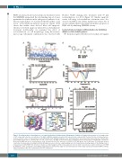
Figure 2. A natural product, isobavachalcone, is a newly identified direct dihydroorotate dehydrogenase inhibitor. (A) Graphical presentation of screening results of 337 compounds tested at a concentration of 10 μM in a DHODH enzymatic assay. Each dot represents one compound. (B) Chemical structure of isobavachalcone. (C) Dose-response curves of isobavachalcone and leflunomide in the DHODH enzymatic assay. (D) A thermofluor assay shows that isobavachalcone robustly stabi- lizes DHODH and produces a thermal shift over 14°C (ratio 1:10). (E) NMR measurement of direct binding between isobavachalcone and DHODH. Carr-Purcell- Meiboom-Gill NMR spectra for isobavachalcone (red), isobavachalcone in the presence of DHODH at 2.5 μM (green) and 5 μM (cyan). (F) Isothermal titration calorimetry of isobavachalcone binding to DHODH. Binding curves were fitted as a single binding event. (G) Computational docking analysis of the binding mode of isobavachalcone with DHODH. The structure is shown as a ribbon diagram and the isobavachalcone molecule (left) is presented as a sphere model based on PDB ID: 4YLW. The amino acid residues surrounding isobavachalcone (yellow sticks, right) are represented by slate sticks. Figure 1G was generated by PyMOL software (https://www.pymol.org/). IBC: isobavachalocone; LEF: leflunomide.
1476
haematologica | 2018; 103(9)
