
Page 75 - 2018_09-Mondo
P. 75
Inhibition of DHODH induces differentiation in AML
DHODH markedly reduced the sensitivity of HL60 cells to isobavachalcone (Figure 3E). Taken together, these findings indicate that isobavachalcone suppresses HL60 cell growth through direct inhibition of DHODH.
After treatment with increasing concentrations of isobavachalcone for 72 h, the percentage of apoptotic cells
A
B
C
D
increased in a dose-dependent manner (Figure 3F,G). To fur- ther investigate the mechanisms underlying isobavachal- cone-induced apoptosis in HL60 cells, we performed a western blot assay to detect the protein marker of apopto- sis. Figure 3H reveals that several protein markers of apop- tosis (cleaved caspase-9, cleaved caspase-3 and cleaved
F
G
EH
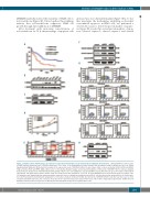
Figure 1. Dihydroorotate dehydrogenase is required for acute myeloid leukemia cells to maintain their malignant characteristics.. (A) Kaplan-Meier survival curves for AML patients divided by level of DHODH expression. The P-value of the Kaplan-Meier survival analysis was determined using a log-rank test (see the Online Supplementary Methods). (B) Western blot analysis of the expression levels of DHODH in different cancer types. SiHa: cervical carcinoma; H1299, A549 and H446: lung carcinoma; HL60 and THP1: AML; Jurkat: acute T-cell leukemia; HepG2: hepatic carcinoma; U251: glioma. (C) Knockout of DHODH in HL60 cells was analyzed by western blot. (D) Knockout of DHODH impaired the growth of HL60 cells. Cell viability was evaluated by MTS assay at 24 h intervals up to 96 h in three independent experiments. The graph represents the means ± SD. The Student t-test was performed, **P<0.01. (E and F) DHODH knockout resulted in apoptosis of HL60 cells. Cell apoptosis was analyzed by flow cytometry and the expression levels of apoptosis-related proteins in HL60 cells was detected by western blot at 96 h after infec- tion. (G) Flow cytometry demonstrated upregulation of cell surface markers CD14 and CD11b after knockout of DHODH in HL60 cells whereas there was no effect on CD33 and CD34 expression. The cells were measured at 96 h after infection. Data represent the mean ± SD of three independent experiments. (H) Knockout of DHODH resulted in reduced expression of MYC protein and upregulated expression of p21 in HL60 cells.
haematologica | 2018; 103(9)
1475
