
Page 126 - Haematologica August 2018
P. 126
1362
J. Muller et al.
ABCE
D
F
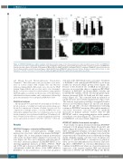
Figure 2. OTSSP167 inhibits bone matrix resorption. A+B) Representative images of Von Kossa-stained bone matrix resorption assays of huOC and RAW264.7- derived osteoclast cultures continuously treated with a range of OTSSP167 concentrations. C+D) Quantification of the matrix resorption area in huOC and RAW264.7- derived osteoclast cultures. E) Real-time PCR analysis of MELK, IRF8, DC-STAMP and NFATc1 following OTSSP167 treatment of RAW264.7-derived osteoclast cul- tures. F) Representative confocal microscopy images of phalloidin-FITC-stained RAW264.7-derived osteoclast cultures. All data are represented as mean +/- stan- dard error. *: P<0.05, **: P<0.01, ***: P<0.001. For clarity, only significant differences with vehicle-treated cultures are shown.
and Mineral Research Histomorphometry Nomenclature Committee.22 Osteoblast surface and osteoid surface were meas- ured on sections stained with toluidine blue and Masson’s trichrome (Sigma-Aldrich). Osteoclasts were detected by TRAP staining (Sigma-Aldrich) and osteoclast surface was determined. Briefly, sections were stained for acid phosphatase using naphthol ASTR phosphate as substrate in the presence of 50 mM tartrate with hexazotised pararosaniline, and counterstained with methyl green. Measurements were performed in NDP.View 2.6.13.
Statistical analyses
All experiments were performed at least in triplicate. Results are shown as means +/- standard error and representative images are shown. For comparisons of 2 means, a Student t-test was used. For comparisons of multiple means, a one-way ANOVA was used, followed by a Dunnett’s post-hoc test or Tukey’s post-hoc test. All statistical analyses were performed with Prism 5 (Graphpad soft- ware). P-values below 0.05 were considered significant and P-val- ues are represented as follows: *=P<0.05, **=P<0.01, ***=P<0.001.
Results
OTSSP167 hampers osteoclast differentiation
During normal osteoclastogenesis, MELK levels increased in osteoclasts cultured from murine RAW264.7 cells (Figure 1A) or from primary human mononuclear cells (results not shown) when compared to monocyte cul- tures (Figure 1A). We could confirm the earlier described15 osteoclast activation pathway (EZH2-IRF8-NFATC1) by showing significant increases in EZH2, NFATC1, TRAP and CTSK and a decrease in IRF8 mRNA (Figure 1A). We subsequently studied the effects of the MELK inhibitor OTSSP167 on the proliferation of osteoclast progenitor
cells and on the differentiation into osteoclasts. Treatment of RAW264.7 cells with 25 nM OTSSP167 for 24 hours resulted in decreased MELK protein levels (Figure 1B). Because of the described role of MELK in cell cycle pro- gression, we assessed the effect of continuous OTSSP167 treatment on RAW264.7 and human PBMC viability, and found that the viability of both osteoclast progenitor pop- ulations decreased (IC50: 12.8 nM and 43.2 nM, respec- tively) (Figure 1C). This coincided with an induction of G2/M cell cycle arrest (Online Supplementary Figure S1A). The decrease in progenitor viability corresponded with a decrease in both human (Figure 1D and 1F) and murine (Figure 1E and 1H) osteoclast differentiation following continuous OTSSP167 treatment. Although the number of osteoclasts decreased, osteoclast size was markedly increased in human cultures treated with 10 nM OTSSP167, with a corresponding increase in the number of nuclei per osteoclasts (Figure 1G). This was not the case for RAW264.7 osteoclast cultures (Figure 1I).
OTSSP167 inhibits bone matrix resorption
Bone matrix resorption decreased following OTSSP167 treatment of both primary human (Figure 2A and 2C) and murine (Figure 2B and 2D) osteoclast cultures. Of note, this effect was apparent in human osteoclasts treated with 10 nM OTSSP167, suggesting that the enlarged osteoclasts that are present at this concentration are not efficiently resorbing matrix. Similarly, murine osteoclast bone resorption was inhibited at 1 nM OTSSP167, a concentration at which no effect on osteoclast differentiation was observed. The block- ing effect of OTSSP167 on osteoclast differentiation was confirmed at mRNA level, where we observed an increased expression of the negative regulator IRF8 and decreased expression of NFATC1 and DCSTAMP (Figure 2E). On the
haematologica | 2018; 103(8)
