
Page 128 - Haematologica August 2018
P. 128
1364
J. Muller et al.
pared to healthy controls was completely prevented fol- lowing treatment with OTSSP167 (Figure 5C). This was due to an increase in the number of trabeculae (Tb.N) (Figure 5D) and a decrease in trabecular separation (Tb.Sp) (Online Supplementary Figure S1F). As a result, trabecular connectivity density (Conn.Dn) was similar to healthy mice in OTSSP167-treated MM-bearing mice (Online
Supplementary Figure S1G). Trabecular thickness was not affected in MM-bearing mice (Online Supplementary Figure S1H). Importantly, the observed prevention of MMBD in these mice does not appear to be solely due to a decreased tumor load following OTSSP167 treatment, as MMBD was prevented at a dose (7.5 mg/kg/2d) which had no effect on tumor load (Figure 5E).
ABC
DEF
G
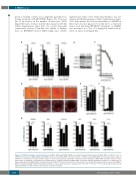
Figure 4. OTSSP167 stimulates osteoblast function. A) MELK, EZH2 and FOXM1 mRNA levels during osteoblast differentiation of BMSC-TERT cells. B) The effect of OTSSP167 treatment on MELK, EZH2 and FOXM1 protein levels in BMSC-TERT osteoblast cultures. C) MTT assay on BMSC-TERT cells incubated with a range of OTSSP167 concentrations. D) Representative images of sirius red (top panels) and alizarin red (bottom panels) stainings of BMSC-TERT osteoblast cultures treated with a range of OTSSP167 concentrations. E) Quantification of collagen deposition by BMSC-TERT osteoblasts following OTSSP167 treatment. F) Quantification of matrix mineralization by BMSC-TERT osteoblasts following OTSSP167 treatment. G) Real-time PCR analysis of osteoblast differentiation marker expression by BMSC- TERT osteoblasts following OTSSP167 treatment. All data are represented as mean +/- standard error. *:P<0.05, **:P<0.01, ***:P<0.001. Except for panel A, only significant differences with vehicle-treated cultures are shown for clarity.
haematologica | 2018; 103(8)
