
Page 125 - Haematologica August 2018
P. 125
OTSSP167 has activity in myeloma bone disease
Treatment of myeloma-bearing mice with OTSSP167
We used the C57BL/KaLwRij 5TGM.1 mouse model21 to assess the effect of OTSSP167 on the development of MMBD. Nine- week-old female mice were injected i.v. with 5.0 x 10⁵ 5TGM.1GFP+ cells and OTSSP167 or vehicle solution was admin- istered by oral gavage at different dose levels. For every cohort, mice were randomly divided into a naive group (no MM, n=5), a vehicle group (MM + vehicle, n=10) and a treated group (MM + OTSSP167, n=10). When mice showed signs of active myeloma, i.e., paraplegia, all mice within a cohort were sacrificed. Bone mar- row plasmocytosis was determined by flushing the bones of one leg followed by flow cytometry on a FACSCanto flow cytometer (BD Biosciences). The femur and tibia of the opposite leg were
defleshed and stored in 70% EtOH. Ethical approval was obtained for all mouse experiments (ULg license no. 1336).
Bone histomorphometry
Femurs were embedded in methylmethacrylate. All parameters were recorded as recommended by the American Society for Bone
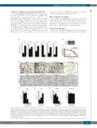
Figure 1. OTSSP167 hampers osteoclast differentiation. A) mRNA levels of MELK, EZH2, FOXM1 and relevant transcription factors during osteoclastogenesis in murine cultures, MO: monocytes; OC: osteoclasts. B) The effect of OTSSP167 treatment on MELK protein expression in RAW264.7 cultures. C) MTT assay on PBMCs and RAW264.7 cells incubated with a range of OTSSP167 concentrations. D+E) Representative images of TRAP-stained human (huOC) and RAW264.7-derived osteo- clast cultures continuously treated with a range of OTSSP167 concentrations. F) Quantification of osteoclast numbers per field of view (N.OC/FOV) in huOC cultures. G) Number of nuclei per osteoclast in huOC cultures. H) Quantification of N.OC/FOV in RAW264.7-derived cultures. I) Number of nuclei per osteoclast in RAW264.7- derived cultures. All data are represented as mean +/- standard error. *: P<0.05, **: P<0.01, ***: P<0.001. For clarity, only significant differences with vehicle- treated cultures are shown.
Micro-computed tomography
Micro-computed tomography (μCT) was performed on distal femurs with the Skyscan 1172 system (Bruker), as described pre- viously.19 3D models of bones were generated using CTVol soft- ware (Bruker). The number of cortical perforations > 50 μm in diameter was counted blinded on reconstructed images.
AB
D
E
FGHI
C
haematologica | 2018; 103(8)
1361
