
Page 106 - Haematologica August 2018
P. 106
C. Melani et al.
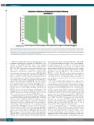
Figure 3. Tumor reduction by end-of-treatment CT. Reduction of the bi-dimensional product of the largest mediastinal mass for the 89 patients with complete tumor measurements by EOT CT. All patients had reduction in tumor bi-dimensional product and there was no relationship between EOT tumor reduction and EOT FDG- PET Deauville score. No difference in tumor reduction was demonstrated between patients with (N=6, red arrows) and without (N=83) treatment failure; Median reduction 92% (range, 65-99) vs. 93% (range, 62-100), respectively.
1342
Our results indicate that very few patients require post- treatment radiotherapy following DA-EPOCH-R, irre- spective of their EOT FDG-PET scans. These findings provide substantial evidence that patients with negative EOT FDG-PET scans rarely recur and are unlikely to ben- efit from additional mediastinal radiotherapy. Furthermore, they provide evidence for our initial obser- vation that most patients with a positive EOT FDG-PET achieve long-term remission following DA-EPOCH-R and, as a group, would not benefit from empirical consol- idation radiotherapy. Indeed, the discrepancy between our findings that routine consolidation radiotherapy is unnecessary following DA-EPOCH-R, and the accepted need for post-treatment radiotherapy in patients with pos- itive EOT FDG-PET scans following R-CHOP has led to uncertainty. Unfortunately, it is not uncommon for patients with a positive EOT FDG-PET following DA- EPOCH-R to receive post-treatment radiotherapy. Such an approach in our study would have resulted in 31% (25/80) of patients receiving radiotherapy, most of whom (80%) were already cured with DA-EPOCH-R alone.
A clinically important aspect of our study is distinguish- ing treatment failures following DA-EPOCH-R. Given the worse outcome of PMBCL compared to DLBCL with sal- vage therapy,29 early recognition of patients with persist- ent disease is critical to optimize the curative potential of radiotherapy while averting its use in patients already cured with DA-EPOCH-R. We first looked at tumor mass reduction based on EOT CT, and observed no predictive value on outcome or any relationship with EOT FDG-PET. We also assessed the ability of single EOT and serial FDG- PET imaging to detect treatment failure. Following DA- EPOCH-R, 69% of patients had a negative EOT FDG-PET. Notably, 98% of these patients never progressed, indicat-
ing such patients rarely require radiotherapy. Among the 31% of patients with positive EOT scans, only 5 ultimate- ly had treatment failure of which 4 occurred in patients with Deauville 5 scans. These results are consistent with the prospective IELSG-26 study, which revealed a signifi- cantly worse outcome in patients with Deauville 4-5 EOT FDG-PET scans (5-yr. PFS 68% vs. 99%, P<0.0001; 5-yr. OS 83% vs. 100%, P=0.003), with the greatest number of treatment failures in Deauville 5 patients.22 In contrast to our study, however, variable chemoimmunotherapy was used and most patients (89%) received consolidation radiotherapy.
We found serial FDG-PET imaging to be a highly effec- tive strategy to distinguish persistent disease from post- treatment inflammatory changes. Linear regression analy- sis in 17 non-progressing patients with a positive EOT FDG-PET and serial imaging showed an overall decrease in SUVmax across serial scans. In contrast, serial FDG-PET imaging in 5 treatment failures with serial scans showed an increase in SUVmax that was statistically greater than patients who never progressed, regardless of EOT FDG- PET response (P=0.011 and P=0.0037 for positive and neg- ative EOT FDG-PET non-progressors, respectively). Overall, use of serial FDG-PET imaging effectively reduced radiotherapy from a potential 31% (25/80) of patients with a positive EOT FDG-PET scan to only 5 (5%) patients with confirmed treatment failure.
We also explored the use of quantitative FDG-PET parameters (i.e., MTV and TLG) to assess if they improved upon SUVmax in identification of treatment failures. These methods were limited by the overall low volume of dis- ease following therapy as well as inability to exclude non- malignant causes of FDG uptake, resulting in a wide vari- ability in value between patients. Although these param-
haematologica | 2018; 103(8)
