
Page 107 - Haematologica August 2018
P. 107
Serial FDG-PET Imaging in PMBCL
AB
CD
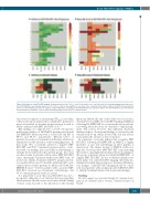
Figure 4. Evolution of serial FDG-PET imaging. Heatmap depiction of (A). SUVmax, and (B). Deauville score, over time in the 20 non-progressing patients with a pos- itive EOT FDG-PET scan. Heatmap depiction of (C). SUVmax, and (D). Deauville score, over time in the 6 patients with treatment failure. FDG-PET scans performed prior to the EOT FDG-PET are listed as negative numbers with those following the EOT FDG-PET listed as positive numbers. The EOT FDG-PET scan is bordered by black dashed lines. FDG-PET scans performed following salvage intervention are shaded in black.
eters were not superior to monitoring SUVmax in our study, other recent reports indicate these quantitative parameters may be beneficial for baseline prognostication as well as when combined with EOT Deauville score.30,31
Our findings are supported by a recent retrospective multi-center analysis of 156 PMBCL patients treated with DA-EPOCH-R which reported a 3-year EFS and OS of 85.9% and 95.4%, respectively.32 Overall, 14.9% of patients received post-treatment radiotherapy, which was administered at the discretion of the treating physician. In that study, 75% of patients achieved a negative EOT FDG-PET and 95.4% remained progression-free, consis- tent with our findings that consolidation radiotherapy is virtually never indicated in this patient group. Less clear are their results in patients with positive EOT FDG-PET scans. Among the 31 patients with positive EOT scans, 19 received no further treatment with 68% progression-free at a median follow up of 17 months, indicating that a sub- stantial subset of these patients are likely cured with DA- EPOCH-R alone.32 Twelve patients with a positive EOT FDG-PET received post-treatment radiotherapy and 33.3% remain progression-free at 2 years.
It is important to note that serial FDG-PET was not a prospective endpoint of our trial and decisions regarding which patients should receive serial scans and the timing of those scans was left to the discretion of the treating
physician. Indeed, the aim of this study was to provide a descriptive look at EOT and serial PET imaging in PMBCL following DA-EPOCH-R as it occurs in the real-world clin- ical setting, where decisions are often left to clinical judge- ment. The notion, however, that physician discretion influenced these observational findings is obviated by the extended follow up, which showed who did and did not recur and by the absence of late recurrences.
In conclusion, our results indicate that a negative EOT FDG-PET following DA-EPOCH-R in PMBCL is highly predictive of cure and radiotherapy in these patients is unnecessary. The unique biology of PMBCL results in a high rate of false-positive EOT FDG-PET scans indicating the need for a paradigm shift in clinical decision making for this group of patients when receiving DA-EPOCH-R. A singular EOT FDG-PET did not accurately identify treat- ment failure but serial FDG-PET imaging effectively dis- criminated residual disease from post-treatment inflamma- tory changes. Serial FDG-PET imaging should be consid- ered in all patients with an initial positive EOT FDG-PET to identify treatment failures that require radiotherapy.
Funding
Research support was provided through the intramural pro- gram of the National Cancer Institute, National Institutes of Health.
haematologica | 2018; 103(8)
1343
