
Page 73 - Haematologica June
P. 73
Effects of HC and IFN-α on PV RBC adhesion
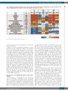
Table 1. ITRAQ ratios of proteins with increased (i) or decreased (d) expression at the membrane of polycythemia vera (PV) red blood cells, and effect of hydroxycarbamide (HC) treatment. Proteins from endoplasmic reticulum are in bold.
CT: control; PVHC: blood sample from HC-treated patient.
37 proteins (ratio ≥1.3) and downregulation of 16 proteins (ratio ≤0.7) (Table 2).
Expression of Calr was diminished during HC treatment but did not reach control levels (Table 1). We analyzed Calr by western blot in RBC ghosts from a total of 4 pre- post patients, including the 3 patients of the proteomics study, and found that its expression was decreased during the treatment without reaching significance (Figure 1A), confirming the proteomics results. This was probably due to the variability and relatively short duration of the treat- ment in this group (mean duration 1.4 years, range 0.5-3 years). Therefore, we tested for the presence and expres- sion level of Calr by western blot in a group of 11 patients treated with HC for a longer period of time (mean dura- tion 5.9 years, range 0.5-20 years), a group of 19 untreated (UT) patients (i.e. with no cytotoxic antiproliferative treat- ment), and a group of 11 healthy donors (Figure 1B). We found no significant difference between the HC and healthy donor groups indicating that long-term HC treat- ment restores the expression of Calr. Similarly, we inves- tigated Calr in a group of 7 patients treated with IFN and found no difference with the healthy donors group, strongly suggesting that IFN restores normal expression of Calr in PV RBCs (Figure 1B).
Overexpression of Lu/BCAM and CD147 in HC-treated patients
In the group of membrane proteins abnormally expressed in PV RBCs,19 Lu/BCAM was the only protein whose overexpression was further exacerbated by HC in all 3 patients, with a fold increase comprised between 6.6 and 11.8 (Table 1). Lu/BCAM is a low abundance surface protein that is expressed on a subpopulation of circulating RBCs. To determine whether HC increases the percentage
of Lu/BCAM-positive RBCs or Lu/BCAM expression level per RBC, or both, we performed flow cytometry assays with 18 UT and 17 HC blood samples, using a specific mouse monoclonal anti-Lu/BCAM antibody. The percent- age of Lu/BCAM-expressing cells and the number of Lu/BCAM molecules per RBC, estimated by the mean flu- orescence intensity (MFI) of Lu/BCAM-positive RBCs, were significantly higher in the HC group (median 94.35%, MFI=4570) than in the UT group (median 73%, MFI=1819) (P<0.001, Mann-Whitney test) (Figure 2A and B, and Online Supplementary Figure S1A). As Lu/BCAM expression is known to be higher in young (reticulocytes) than mature RBCs,25 we determined the percentage of reticulocytes in all blood samples. There was no signifi- cant difference between both patient groups (mean UT:0.6 ±0.05; HC: 0.9±0.17, P=0.097) (Online Supplementary Figure S1B) indicating that the increase of Lu/BCAM was not due to an HC-induced imbalance between reticulocytes and mature RBCs.
Flow cytometry analysis was conducted with 11 blood samples from IFN-treated patients and no significant dif- ference was found with the UT group (Lu/BCAM-positive RBCs: 81.4% vs. 73%, P=0.3722; MFI: 2479 vs. 1819, P=0.5588, respectively) (Figure 2A and B), indicating that IFN did not influence Lu/BCAM protein expression.
In order to explore the potential effect of HC and IFN on the expression of other erythroid adhesion proteins, we performed flow cytometry analysis of four additional adhesion markers: CD44, CD47, CD147 and CD242. All 4 markers are expressed on almost 100% of circulating RBCs and there was no effect of HC or IFN on this per- centage (data not shown). Likewise, there was no effect of either HC or IFN treatment on the expression level per RBC of CD44, CD47 and CD242, estimated by the MFI,
haematologica | 2018; 103(6)
975
