
Page 274 - Haematologica Vol. 107 - September 2022
P. 274
LETTER TO THE EDITOR
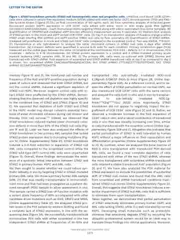
Figure 1. STAG1 knockdown perturbs STAG2 null hematopoietic stem and progenitor cells. Umbilical cord blood (UCB) CD34+ cells were cultured in serum-free expansion medium (SFEM) added with stem cell factor (SCF), thrombopoietin (TPO) and FMS- like tyrosine kinase 3 ligand (FLT3L) at final concentration of 100 ng/mL each. (A) Flow cytometry analysis of enhanced green fluorescent protein (eGFP) expression in UCB CD34+ cells edited with either mock or with single guide RNA (sgRNA) (UCUGGUCCAAACCGAAUGAA) - Cas9 ribonucleoproteins targeting STAG2 along with adeno-associated virus donor template. (B) Quantification of CRISPR/Cas9-mediated eGFP knockin efficiency measurement across 3 replicates. (C) Western blot analysis of STAG2 protein in the mock and eGFP sorted UCB CD34+ cells. (D) Day 5 co-transduction analysis of Kusabira orange positive STAG1 small interfing RNA (shRNA) and eGFP-positive STAG2 null cells by flow cytometry. (E) Quantification of STAG1 shRNA- mediated cell proliferation in mock and STAG2 null CD34+ cells compared to the scrambled control. Two-way ANOVA, **P<0.01. (F) Fluorescence in situ hybridization to analyze the sister chromatid cohesin in STAG2 null cells 3 days after shRNA transduction. (G) Cohesion defects were quantified in around 8-15 cells for each condition. Primary constriction gaps (PCG) measured are the visible gaps between the sister chromatids at the centromeres; PCG mild - defects in 1-4 chromosomes, PCG moderate - defects in 4-19 chromosomes. (H) STAG2 protein expression in a control and 2 STAG2-mutated primary acute myeloid leukemia (AML) samples (AML 16 and 94). (I) The primary AML cells were co-cultured with OP9M2 Stroma and transduced with STAG1 shRNA. Fold expansion of scrambled and STAG1 shRNA transduced cells at day 5 as compared to day 2 is shown. Scr: scrambled shRNA CAACAAGATGAAGAGCACCAA; Sh1: STAG1 shRNA1 CTTCAGCCTTTGGTGTTCAAT; Sh2: STAG1 shRNA2 GCCAATGAAAGGTTGGAGTTA.
mentaryFigure1CandD).Wemonitoredcellnumberand frequency of the KuO and GFP-positive population during 1 week of culture and observed that both STAG1 shRNA, but not the control shRNA, induced a significant depletion of STAG2 null HSPC. Moreover, isogenic control cells with in- tact STAG2 were unaffected by the STAG1 shRNA, demon- stratingthattheobservedcelldepletionwashighlyspecific to the combined loss of STAG1 and STAG2 (Figure 1D and E). We reasoned that depletion of both STAG1 and STAG2 in the HSPC model may disrupt cohesin’s essential func- tions of sister chromatid cohesion during cell division and thereby limit cell survival.11,12 Indeed, we observed that STAG1 knockdown induced marked sister chromatid cohe- sion defects in more than 50% of the STAG2 null cells (Fig- ure 1F and G). Later we have also analyzed the effects of STAG1 knockdown in two primary AML samples that lacked STAG2 protein expression due to truncating mutations (Fig- ure 1H; Online Supplementary Table S1). STAG1 knockdown induced a 2-5-fold reduction in expansion of STAG2 null AML cells compared to the scrambled control while the STAG2 wild-type (WT) control AML cells were unperturbed (Figure 1I). Overall, these findings demonstrate the exist- ence of a synthetic lethal interaction between STAG1 and STAG2 in primary human HSPC and AML cells.
We then sought to assess whether we could induce syn- thetic lethality in vivo by targeting STAG1 in STAG2-mutated primary AML cells. We chose a primary human AML sample (AML 21) that was readily transplantable in immunodefi- cient mice and that had been propagated as a patient-de- rived xenograft (PDX) sample to allow assessment in vivo. This sample carried a STAG2 loss-of-function mutation with a variant allele frequency of 89% accompanied with other candidate driver mutations such as IDH2, SRSF2 and NRAS. (Online Supplementary Table S1). We analyzed STAG2 pro- tein levels in the PDX sample by western blotting and found a complete lack of STAG2 expression, in line with the se- quencing data (Figure 2A). We successfully transduced bulk mononuclear PDX cells with either scrambled or the two independent STAG1 shRNA at frequencies of 30-60% and
transplanted into sub-lethally irradiated NOD-scid IL2Rgnull-3/GM/SF (NSG-S) mice (Figure 2B; Online Sup- plementary Figure S2A). As a reference control, and to as- sess the effect of STAG1 perturbation on normal HSPC, we also transduced UCB CD34+ cells with the same vectors and assayed the cells both in vitro and in vivo by transplan- tation to sub-lethally irradiated NOD.Cg- PrkdcscidIl2rgtm1Wjl/SzJ (NSG) mice. Importantly, STAG1 knockdown did not appear to negatively impact the en- graftment of UCB CD34+ cells (Figure 2C and D). Rather, we observed a moderate increase in the fold expansion of CD34+ cells in vitro, and a robust contribution of transduced cells in vivo that was steadily increasing over time, similar to cells transduced with the scramble control (Online Sup- plementary Figure S2B and C). Altogether this indicates that partial perturbation of STAG1 is well tolerated by human HSPC without a major influence on their repopulation and differentiation properties (Online Supplementary Figure 2D to H). By contrast, when we analyzed the bone marrow of NSG-S mice transplanted with transduced PDX-derived AML cells, we found a near complete depletion of cells transduced with either of the two STAG1 shRNA, whereas the mice transplanted with scrambled shRNA transduced cells retained a stable transduced KuO+ population (Figure 2E and F). We have also analyzed the PDX material for STAG2 expression to exclude the possibilities of a potential drift of STAG2 null clones and found that the AML cells from scrambled and shRNA transduced conditions main- tained STAG2 null clones (Online Supplementary Figure 2I). Overall, this suggests that STAG1 knockdown induces a se- lective impairment of STAG2 null AML cells that is sufficient to eliminate them upon transplantation.
Taken together, we demonstrate that partial perturbation of STAG1 selectively eliminates primary human HSPC and AML cells lacking STAG2, while it is well tolerated by normal HSPC. Developing small molecules such as proteolytic chimeras that selectively degrade STAG1 by recruiting the ubiquitin-proteasomal system would be an ideal way to translate these findings into clinical applications. Moreover,
Haematologica | 107 September 2022
2273
