
Page 252 - Haematologica Vol. 107 - September 2022
P. 252
LETTER TO THE EDITOR
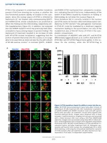
ETV6 in the cytoplasm to understand whether mutations prevent ETV6 from entering the nucleus or whether the non-functional proteins rapidly re-localize to the cyto- plasm. Since the nuclear export of ETV6 is inhibited by leptomycin B,12 we treated cells overexpressing Q347P- and R399H-ETV6, whose substitutions are predicted to affect the folding and the DNA binding, respectively (On- line Supplementary Figure S1). In addition, we analyzed also the W380R mutation which has previously been dem- onstrated to have a strong impact on protein folding.6 The leptomycin B treatment resulted in an increase of cells with WT-ETV6 nuclear localization, suggesting that the portion of protein that enter into the nucleus is no longer able to be exported, as observed also for the p65 subunit of NF-kB used as control.13 In contrast, Q347P-, W380R-
and R399H-ETV6 maintained their cytoplasmic localiza- tion, indicating that M-ETV6 forms, independently of the extent of the defect caused by mutations on folding or DNA binding, do not enter the nucleus (Figure 2).
Since mutations fail to correctly localize in the nucleus and ETV6 exerts its function after homodimerization me- diated by its PNT domain,14 the pathogenetic mechanism in ETV6-RT could be mediated by a dominant negative mechanism leading to the accumulation not only of the mutated but also of the WT forms of ETV6 in the cyto- plasmic compartment.
Co-transfection of HEK293T cells with WT- and M-ETV6 differentially tagged allowed us to confirm that both WT- ETV6-Myc and WT-ETV6-Flag proteins gather in the nu- cleus. On the contrary, when the WT-ETV6-Flag is
AB
Figure 2. ETV6 mutations impair its ability to enter into the nu- cleus. (A) ETV6 immunostaining in HeLa cells after 4 hours of Leptomycin B (LB) treatment (50 nM). Subcellular localization of Q347P, W380R and R399H mutation (red) and p65 (green) before (NT) and after LB treatment. Nuclei were marked with Hoechst staining (blue). Images were obtained with a Zeiss Axioplan 2 epifluorescence imaging microscope and acquired with a Zeiss Axiocam 506 color using a 40X Plan-NEOFLUAR objective. Images were processed using Zeiss ZEN 3.1 (blue edi- tion), while brightness and contrast were adjusted using Adobe Photoshop 2020. (B) Cell counts basing on ETV6 subcellular lo- calization before and after treatment with LB. Histogram rep- resents cell fraction with nuclear (N), cytoplasmatic (C) or both (N/C) staining, confirming the intracellular distribution variation of ETV6 variants after LB treatment. All data reported are rep- resentative of at least 3 independent experiments.
Haematologica | 107 September 2022
2251
