
Page 251 - Haematologica Vol. 107 - September 2022
P. 251
LETTER TO THE EDITOR
AB
C
D
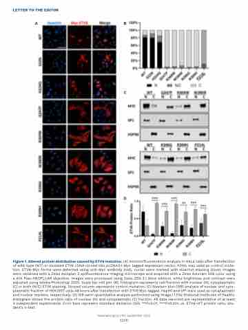
Figure 1. Altered protein distribution caused by ETV6 mutation. (A) Immunofluorescence analysis in HeLa cells after transfection of wild-type (WT) or mutated ETV6 cDNA cloned into pcDNA3.1-Myc tagged expression vector. P214L was used as control muta- tion. ETV6-Myc forms were detected using anti-Myc antibody (red), nuclei were marked with Hoechst staining (blue). Images were obtained with a Zeiss Axioplan 2 epifluorescence imaging microscope and acquired with a Zeiss Axiocam 506 color using a 40X Plan-NEOFLUAR objective. Images were processed using Zeiss ZEN 3.1 (blue edition), while brightness and contrast were adjusted using Adobe Photoshop 2020. Scale bar =50 μm. (B). Histogram represents cell fraction with nuclear (N), cytoplasmatic (C) or both (N/C) ETV6 staining. Striped column represents control mutation. (C) Western blot (WB) analysis of nuclear and cyto- plasmatic fraction of HEK293T cells 48 hours after transfection with ETV6 Myc-tagged. Hsp90 and SP1 were used as cytoplasmatic and nuclear markers, respectively. (D) WB semi-quantitative analysis performed using ImageJ 1.53e (National Institutes of Health). Histogram shows the protein ratio of nuclear (N) and cytoplasmatic (C) fraction. All data reported are representative of at least 3 independent experiments. Error bars represent standard deviation (SD). **P<0.01, ***P<0.001, vs. ETV6-WT protein ratio, Stu- dent’s t-test.
Haematologica | 107 September 2022
2250
