
Page 202 - Haematologica Vol. 107 - September 2022
P. 202
ARTICLE - ITP antibody predicts desialylation and apoptosis S.S. Zheng et al.
Table 2. Relative effects of anti-GPIIb/IIIa and anti-GPIb/IX antibodies on platelet desialylation and apoptosis.
treatment for a larger proportion of ITP patients as anti- GPIIb/IIIa antibody is a more common antibody than anti- GPIba antibody.
Discussion
ITP is a heterogenous disease with multiple proposed mechanisms. Potential therapeutic advances require more detailed understanding of the means that lead to platelet destruction. As such, we sought to examine platelet de- sialylation and apoptosis as contributors to thrombo- cytopenia in ITP. We studied 61 ITP patient sera and examined the presence of APA as a predictor for these two processes. Antibody specificity was interrogated, specifi- cally, anti-GPIb/IX, anti-GPIIb/IIIa and GPV antibodies. GPIa was not examined as isolated anti-GPIa/IIa antibody posi- tivity has not been reported in recent literature.43,47 We demonstrated that the presence of APA in ITP patients’ sera is associated with platelet desialylation in our patient population. Although desialylation was initially thought to be induced by anti-GPIb/IX antibodies in ITP,28 here we found that enhanced neuraminidase expression was ob- served in the majority of our patient cohort with detect- able APA. This supports recent studies which reported that the loss of sialic acid is a more frequent finding in ITP than previously thought.36,48 Furthermore, in a murine model of ITP utilising patients’ anti-GPIIb/IIIa antibodies and human platelets, we found solid and reproducible36 evidence to support the use of neuraminidase inhibitors as potential new therapeutics for ITP.
The status of whether the patients have detectable ITP antibodies also influences the degree of platelet apopto- sis. Sera with ITP antibodies induced significantly greater loss of ΔΨm compared to the controls, which was not ob- served in the antibody-negative group. As we and others have found that desialylation depends on FcR activity in
Effect
Anti-GPIIb/IIIa sera
Anti-GPIb/IX sera
Positive NEU1 translocation
67%
20%
Significant loss of ΔΨm
33%
80%
GP: glycoprotein; ΔΨm: mitochondrial inner transmembrane poten- tial.
sample also led to decreased ΔΨm compared to controls but was not statistically significant (P=0.15; Kruskal Wallis with Dunn’s multiple comparison), indicating that anti- GPV APA possibly lead to platelet apoptosis. A larger sample size with sole anti-GPV antibody is needed to as- sess its functional effect on platelets.
Oseltamivir protects platelets from GPIIb/IIIa antibody mediated destruction in vivo
Neuraminidase inhibitor oseltamivir has been previously reported to protect platelets from anti-GPIba monoclonal antibody driven platelet destruction in murine studies.28 More recently, we demonstrated oseltamivir’s effect on platelet number preservation in the presence of poly- clonal human anti-GPII/IIIa antibody from a patient with acquired Glanzmann Thrombasthenia.37 Following our findings that anti-GPIIb/IIIa antibodies induce desialyla- tion (Figure 4A), we extended our in vivo experiments to examine other ITP patients with sole anti-GPIIb/IIIa anti- bodies. In order to test whether destruction of human platelets could be prevented in vivo, we treated recipient mice with oseltamivir. As shown in Figure 6, oseltamivir protected human platelets from anti-GPIIb/IIIa antibody- mediated destruction. Therefore, the protective effect of desialylation inhibitors could be generalized to patients with anti-GPIIb/IIIa antibodies. Oseltamivir reduces pla- telet destruction in ITP and is potentially an efficacious
AB
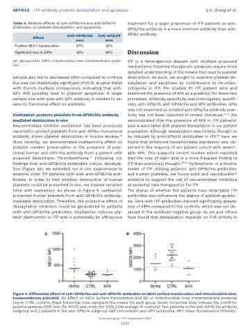
Figure 4. Differential effect of anti-GPIIb/IIIa and anti-GPIb/IX antibodies on NEU1 surface translocation and mitochondrial inner transmembrane potential. (A) Effect on NEU1 surface translocation and (B) on mitochondrial inner transmembrane potential (Δψm). CTRL: control. Black horizontal lines represent the means for each group. Green horizontal lines indicate the cutoff for positive samples (2SD over [for NEU1] and under [for DIOC6] the average of controls). Two patients in the anti-GPIIb/IIIa antibody subgroup and 2 patients in the anti-GPIb/IX subgroup had concomitant anti-GPV antibodies. MFI: mean fluorescence intensity.
Haematologica | 107 September 2022
2201
