
Page 201 - Haematologica Vol. 107 - September 2022
P. 201
ARTICLE - ITP antibody predicts desialylation and apoptosis S.S. Zheng et al.
clinically relevant is that, anti-GPIIb/IIIa auto-antibodies account for the majority of antibody positive cases in our patient population (Figure 1B), which is consistent with a recent report.43 Therefore, further examination into the pathogenesis of anti-GPIIb/IIIa antibody driven ITP is of clinical significance.
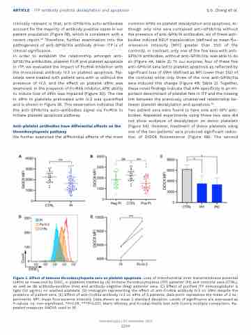
In order to establish the relationship amongst anti- GPIIb/IIIa antibodies, platelet FcγR and platelet apoptosis in ITP, we evaluated the impact of FcγRIIA inhibition with the monoclonal antibody IV.3 on platelet apoptosis. Pla- telets were treated with patient sera with or without the presence of IV.3, and the effect on platelet ΔΨm was examined. In the presence of FcγRIIA inhibitor, APA’ ability to induce loss of ΔΨm was impaired (Figure 3D). The rise in ΔΨm in platelets pretreated with IV.3 was quantified and is shown in Figure 3E. This observation indicates that the anti-GPIIb/IIIa auto-antibodies signal via FcγRIIA to initiate platelet apoptosis pathway.
Anti-platelet antibodies have differential effects on the thrombocytopenic pathway
We further examined the differential effects of the most
common APA’s on platelet desialylation and apoptosis. Al- though only nine sera contained anti-GPIIb/IIIa without the presence of anti-GPIb/IX antibodies, six of these anti- bodies induced NEU1 translocation (defined as mean flu- orescence intensity [MFI] greater than 2SD of the controls). In contrast, only one of the five sera with anti- GPIb/IX antibodies, without anti-GPIIb/IIIa, was able to do so (Figure 4A, table 2). To our surprise, four of these five anti-GPIb/IX sera led to platelet apoptosis as reflected by significant loss of ΔΨm (defined as MFI lower than 2SD of the controls) while only three of the nine anti-GPIIb/IIIa sera induced this change (Figure 4B; Table 2). Together, these novel findings indicate that APA specificity is an im- portant determinant of platelet fate in ITP and the missing link between the previously unobserved relationship be- tween platelet desialylation and apoptosis.36
Two patient sera were found to have sole anti-GPV anti- bodies. Repeated experiments using these two sera did not show evidence of desialylation on donor platelets (Figure 5A). However, treatment of donor platelets using one of the two patients’ sera produced significant reduc- tion of DiOC6 fluorescence (Figure 5B). The second
ABC
DE
Figure 3. Effect of immune thrombocytopenia sera on platelet apoptosis. Loss of mitochondrial inner transmembrane potential (ΔΨm) as measured by DiOC6 in platelets treated by (A) immune thrombocytopenia (ITP) patients’ (Pt) and controls’ sera (CTRL), as well as (B) antibody-positive (Pos) and antibody-negative (Neg) patients’ sera. (C) Effect of purified ITP immunoglobulin G (IgG) (50 μg/mL) on washed platelets. (D) Histogram representing the effect of anti-FcγRIIa antibody IV.3 on ΔΨm despite the presence of patient sera. (E) Effect of anti-FcγRIIa antibody IV.3 on ΔΨm of 3 patients; data point represents the mean of 3 ex- periments. MFI: mean fluorescence intensity. Data shown as mean ± standard deviation. Levels of significance are expressed as P-values. ns: non-significant, *P<0.05, ***P<0.001. Mann Whitney and Kruskal-Wallis test with Dunn’s multiple comparison. Re- peated measures ANOVA used in 3E.
Haematologica | 107 September 2022
2200
