
Page 160 - Haematologica Vol. 107 - September 2022
P. 160
ARTICLE - Use of aqueous humor in VRL management
X. Wang et al.
Early detection of circulating tumor DNA in cerebrospinal fluid in patients with primary vitreoretinal lymphoma may predict parenchymal invasion of the brain
CSF samples were collected from seven patients for whom CSF cytology examinations were negative. Five of the patients were positive for CSF ctDNA and the median concentration of the extracted cfDNA from CSF samples was 1.1 ng/μL (Online Supplementary Table S2). The numbers of alterations in VF/AH and CSF samples are shown in Figure 2B, while the allele frequencies of the shared mutations are shown in Figure 2C. All five pa- tients had significantly higher allele frequencies for the shared mutations in AH and/or VF samples than in CSF ctDNA. It is also of note that patients V1 and V10, who had synchronous PCNSL confirmed by PET/CT examin- ations, harbored a higher number of mutations detected only in CSF samples with significantly higher allele fre- quencies (P<0.01) than those of the other three patients (V5, V6, and V15). Notably, brain involvement developed in patients V5 and V6 after 9 and 19 months, respectively. Thus, positive CSF ctDNA detection might be an early in- dicator of brain involvement with a subset of alterations in patients with primary VRL.
Dynamic profiling of circulating tumor DNA in aqueous humor or vitreous fluid samples to monitor response to treatment
Of the seven VRL patients who received systemic treat- ment, five underwent a series of dynamic AH/VF sampling (4 underwent serial AH sampling and 1 underwent serial VF sampling) (Figure 3, Online Supplementary Table S4). AH or VF samples were collected for ctDNA detection through- out treatment to dynamically monitor the therapeutic re-
sponses. In addition, the IL-10 levels in AH/VF samples were also analyzed to assist with the response evaluation of VRL patients. For patients V5 and V6, the changes of IL- 10 levels were highly concordant with the allele frequencies of AH/VF ctDNA (Figure 3A, B). For patient V15, all four IL- 10 tests were performed within the interval of two rounds of AH ctDNA sequencing, which provided limited informa- tion about the correlation between the IL-10 levels and ctDNA allele frequencies (Figure 3C). The remaining two pa- tients (V2 and V3) with extremely high levels of IL-10 at baseline showed dramatic decreases in post-treatment IL- 10 levels and AH ctDNA allele frequencies (Figure 3D, E). These findings imply that AH/VF ctDNA profiling could as- sist in the evaluation of treatment efficacy, in addition to the use of IL-10 tests and eye examinations.
Comparisons of mutational landscape between patients with vitreoretinal lymphoma and primary central nervous system lymphoma
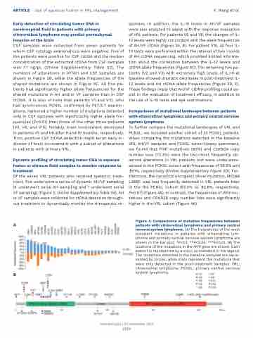
To further compare the mutational landscapes of VRL and PCNSL, we included another cohort of 23 PCNSL patients. When comparing the mutations detected in the baseline VRL AH/VF samples and PCNSL tumor biopsy specimens, we found that PIM1 mutations (80%) and CDKN2A copy number loss (73.3%) were the two most frequently ob- served alterations in VRL patients, but were underrepre- sented in the PCNSL cohort with frequencies of 56.5% and 39.1%, respectively (Online Supplementary Figure S3). Fur- thermore, the canonical oncogenic driver mutation, MYD88 L265P, was less frequently detected in VRL patients than in the the PCNSL cohort (53.3% vs. 82.6%, respectively, P=0.07) (Figure 4A). In contrast, the frequencies of IRF4 mu- tations and CDKN2B copy number loss were significantly higher in the VRL cohort (Figure 4A).
Figure 4. Comparisons of mutation frequencies between A patients with vitreoretinal lymphoma and primary central nervous system lymphoma. (A) The frequencies of the most prevalent mutations in patients with vitreoretinal lym- phoma and primary central nervous system lymphoma are shown in the bar plot. *P<0.1; **P<0.05; ***P<0.01. (B) The locations of the mutations in the IRF4 gene are shown. Each patient is represented by a color, as indicated in the legend. The mutations detected in the baseline samples are repre- sented by circles, while stars represent the mutations that were only detected in the post-treatment samples. VRL: vitreoretinal lymphoma; PCNSL: primary central nervous
B
system lymphoma.
Haematologica | 107 September 2022
2159
