
Page 60 - Haematologica May 2022
P. 60
M. Guipponi et al.
AB
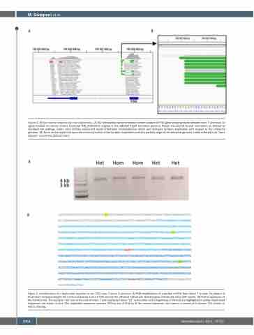
Figure 2. Whole exome sequencing read alignments. (A) IGV (Interactive Genome Viewer) screen capture of FGG gene showing reads between exon 7 and exon 10 (gene located on reverse strand, transcript NM_000509.5) aligned to the GRCh37/hg19 reference genome. Reads are colored by pair orientation as defined by standard IGV settings. Green color defines paired-end reads orientation inconsistencies which can delineate tandem duplication with respect to the reference genome. (B) Zoom on the reads that span the internal junction of the tandem duplication and only partially align to the reference genome (reads referred to as “hard- clipped” at position 155’527’621).
A
B
Figure 3. Identification of a duplicated segment at the FGG exon 7-intron 8 junction. (A) PCR amplification of a portion of FGG from intron 7 to exon 10 yields a 4 kb product corresponding to the normal sequence and a 4.4 kb product for affected individuals. Heterozygous individuals show both bands. (B) Partial sequence of the 4.4 kb band. The acceptor “AG” site at the end of intron 7 and duplicated donor “GT” splice sites at the beginning of intron 8 are highlighted in yellow. Duplicated sequences are shown in blue. The duplicated sequence contains 403 bp out of 404 bp of the normal sequence, one base in a stretch of 4 intronic “A”s (shown in red) is missing.
1068
haematologica | 2022; 107(5)
