
Page 116 - 2022_03-Haematologica-web
P. 116
S.E. Ward et al.
lylated VWF, pdVWF was treated ex vivo with α2-3 neu- raminidase to remove α2-3 linked sialylation from O-gly- cans. In vivo clearance studies were then performed in VWF-/- mice in the presence or absence of combined mMGL1 and mMGL2 inhibition. Removal of α2-3 linked sialylation was associated with a marked reduction in VWF half-life compared to that of the wild-type control (Figure 5A). Importantly, however, this enhanced clear- ance was attenuated in the presence of MGL inhibition (Figure 5A). To assess the relative roles of MGL and ASGPR in modulating the pathological, enhanced clear-
A
ance following removal of α2-3 sialylation, in vivo clear- ance studies were also performed in dual VWF-/-Asgr1-/- knockout mice in the presence or absence of combined mMGL1 and mMGL2 inhibition (Figure 5B). Critically, we observed that MGL inhibition was also able to block enhanced clearance of pdVWF after loss of α2-3 sialyla- tion equally effectively in the presence or absence of ASGPR (Figure 5B).
Terminal sialylation on VWF O-glycans can be either α2-3 or α2-6 linked. In contrast, sialylation on VWF N- glycan chains is predominantly α2-6 linked (Figure
BC
D
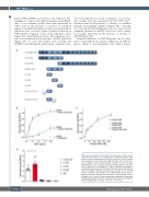
Figure 2. The A domains of von Willebrand factor play a critical role in regulating MGL binding. (A) Schematic of von Willebrand factor (VWF) variants used to characterize the VWF-MGL interaction. All VWF variants were expressed in and purified from HEK293T cells. (B, C) In vitro bind- ing of purified human plasma-derived (pd)VWF (B) and truncated A1A2A3-VWF (C) was assessed using plate binding assays in the pres- ence or absence of 10 mM EDTA or 1 mg/mL ristocetin. (D) Binding to human MGL was assessed for individual A domain proteins (A1-VWF, A2- VWF and A3-VWF). Significant binding was observed for the A1-VWF domain compared with A2-VWF and A3-VWF. Bovine serum albumin (BSA) was used as a negative control. All data are presented as mean ± standard error of mean of three independent experiments. Percentage binding was calculated based on optical density at 450 nm obtained for 100 nM A1A2A3-VWF. *P<0.05, **P<0.01, ***P<0.001.
672
haematologica | 2022; 107(3)
