
Page 107 - 2022_03-Haematologica-web
P. 107
Dendritic cells in myelodysplastic syndromes
A
B
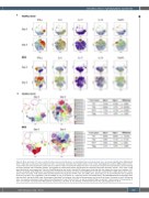
Figure 6. Mass cytometry of T cells co-cultured in the presence of healty donor- or myelodysplastic syndrome-derived slan+ monocytes. Healthy donor (HD)-derived CD4+ T cells were co-cultured in the presence of slan+ non-classical monocytes from healthy donors (n=2) or from myelodysplastic syndrome (MDS) patients (n=2). T cells at the start of the experiment (named “day 0”) as well as T cells co-cultured for 5 days with slan+ non-classical monocytes were stained with a panel consisting of surface markers and intracellular markers, and markers for transcription factors and cytokines and analysed using mass cytometry (CyTOF). First, viable T cells were identified for each experiment. Then the FlowSOM algorithm was used to identify 15 metaclusters containing cells that express the same set of markers. (A) T cells are visualized using viSNE plots. The expression of a selection of markers are shown in the viSNE plots for cultures containing HD- or MDS-derived slan+ mono- cytes at day 0 and day 5. T-cell subsets were identified based on the expression of IFN-γ, Tbet, IL-4, GATA3, IL-17, CD25, CD127, IL-10 and FoxP3 (Th1 were considered to be IFN-γ and Tbet+, IL-17 and GATA3–; Th2 were GATA3+ or IL-4+; Th17 were IL-17+; Tregs were CD127- and FoxP3+CD25+). (B) FlowSOM-identified metaclusters were laid over day 0 and day 5 viSNE maps. Percentages of identified T-cell subsets at the start of the experiment and at day 5 are shown. Compared to day 0, HD-derived slan+ non-classical monocytes mainly induced pro-inflammatory T cells (Th1 and Th17), as well as collateral Tregs. In contrast, T cells cultured in the presence of slan+ non-classical monocytes from MDS patients showed Th1, and above all, Th2 skewing. In HD-derived cultures Th2 cells disappeared at day 5.
haematologica | 2022; 107(3)
663
