
Page 55 - 2022_02-Haematologica-web
P. 55
Cdc42 and aging of human HSC
AB
CDE
FG
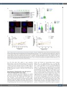
Figure 3. Relative expression of Cdc42 activity in young and aged hematopoietic stem cells. (A) Representative image of western blot. (B) Quantitative expression of relative Cdc42 activity normalized to glyceraldehyde 3-phosphate dehydrogenase (GAPDH). *P= 0.010, if the two points with activity higher than 20 are excluded, then P=0.017; Mann-Whitney test. Bars represent the mean ± standard error of the mean (SEM). nyoung=13; naged=41. Donor age: young =23-39 years (yr), median = 27 yr; aged = 60-82 yr, median = 66 yr. (C) Representative confocal image of Cdc42-GTP expression in hematopoietic stem cells (HSC). Cdc42-GTP quantification of young and aged HSC normalized to (D) DAPI intensity and (E) cell size. ***P=0.0001, **P=0.0012; Mann-Whitney test. Bars = mean ± SEM and nyoung=66; naged=67, from three different donors per cohort. Donor age: young =27-31 years (yr), median =27 yr; aged =63-76 yr, median =76 yr. Scale bar represents 2 mm. (F) Correlation analysis (Spearman) of relative Cdc42 activity and HSC frequency (r=0.4, P= 0.05, n=37) and (G) hematopoietic stem progenitor cell (HSPC) frequency (r= 0.3, P=0.175, n=22), broken grey lines represent 95% Confidence Interval.
tion, the data show that similar to mice, primitive hematopoietic cells from elderly humans show elevated Cdc42 activity. The level of Cdc42 activity in individuals correlates positively with the frequency of HSC, support- ing a possible role for Cdc42 activity in causing the elevat- ed HSC frequency, similar to what has been described in the mouse.8,29
The frequency of hematopoietic stem cells polar for Cdc42 and tubulin declines upon aging
Another established age-related hallmark for murine HSC is the reduction in frequency of cells polar for cytoso- lic polarity proteins like tubulin and Cdc42 (Figure 4A). This “apolarity” of aged murine HSC is a direct conse- quence of the elevated activity of Cdc42 itself in aged HSC9 and likely results in a change in the mode of the division of aged murine HSC.8 We therefore determined the frequency of aged human HSC that showed a polar distribution of
Cdc42 and tubulin by immunofluorescence analyses (Figure 4B to D). Approximately 70% of young HSC showed a polar distribution of Cdc42 while approximately 70% of the aged HSC showed an apolar distribution (Figure 4C). The frequency of aged HSC polar for tubulin was also reduced (Figure 4D). Our findings establish that aged human HSC present with a reduced frequency of cells polar for polarity proteins. Oddly, we did not find a signif- icant association between Cdc42 polarity and tubulin polarity in human HSC (Online Supplementary Figure S5A) suggesting these two parameters might not be directly cor- related as has been shown for murine HSC.9 Nonetheless, we observed a strong negative association between Cdc42 polarity and age (Online Supplementary Figure S5B), imply- ing the frequency of cells that remain polar decreases with increasing age. In order to determine whether the level of Cdc42 activity in human cells might be linked to the fre- quency of cells polar for Cdc42, linear regression analyses
haematologica | 2022; 107(2)
397
