
Page 53 - 2022_02-Haematologica-web
P. 53
Cdc42 and aging of human HSC
ABCD
EF
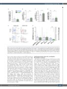
Figure 1. Flow cytometric analysis of different bone marrow populations isolated from young (white) and aged (grey) donors. Populations of interest within the low- density mononuclear cell fraction (MNC) were identified and frequencies of (A) Lin-CD34+ssc low, (B) hematopoietic stem progenitor cells (HSPC), (C) hematopoietic stem cells (HSC) and (D) Lin- cells from young and aged donors were determined. *P<0.04, **P<0.005; Mann-Whitney and t-test with Welch’s correction. Bars rep- resent the mean ± standard error of the mean (SEM). (E) Representative image of common myeloid progenitor/ megakaryocyte–erythroid progenitor (CMP/MEP), multipotent progenitor (MPP) and multipotent lymphoid progenitor (MLP) gates. (F) The frequency of CMP/MEP, MPP and MLP in the Lin- fraction of donors. **P=0.003; Mann-Whitney test. Bars represent the mean ± SEM. 17< nyoung >20; 23< naged >29. Donor age: young =23-39 years (yr), median =27 yr; aged =58-82 yr, median =65 yr. nyoung: number of young donors; naged: number of aged donors.
These results support an increase in the HSPC population with age previously described for the iliac crest.16,23 Our observations further indicate that the age-related increase in frequency is not restricted to a single anatomical site or, in our studies, influenced by sex (Online Supplementary Figure S1B and C). In addition, the frequency of HSC within the Lin-CD34+SSc low population in the sternum was also significantly higher than in the young16 (Online Supplementary Figure S1D). We also found a not yet described increase in the frequency of lineage negative cells within the MNC population in aged donors (Figure 1D). The frequency of common myeloid progenitor/ megakaryocyte–erythroid progenitor (CMP/MEP, Lin- CD34+SSc low CD38+CD90-CD45ra-) and multipotent pro- genitor (MPP, Lin-CD34+SSc low CD38-CD90-CD45ra-) did not change upon aging, while the frequency of immunophenotypic multipotent lymphoid progenitor (MLP, Lin-CD34+SSc low CD38-CD90-/low CD45ra+) with- in the Lin- population decreased significantly (Figure 1E and F). Our results demonstrate and confirm that aging is associated with an increase in the frequency of hematopoietic progenitor and HSC, but with a decrease in the frequency of MLP.
Aged hematopoietic stem cells are delayed in the initiation of division
We next tested whether the age-related increase in the HSC frequency might be linked to an elevated division rate of aged HSC. To this end, we determined the dynam- ics of first or second divisions of individual HSC ex vivo (Figure 2A). In general, BM-derived HSC showed a delayed initiation of division when compared to HSC from cord blood (CB) or HSC mobilized to blood (Online Supplementary Figure S2A and B). Surprisingly, aged HSC actually showed a delay until the first 50% of HSC under- went their first division compared to young HSC (Online Supplementary Figure S2B and C). This delay in initiation of division of aged HSC was still imminent in the presence of a different combination of cytokines (Online Supplementary Figure S2C to E) as well as under normoxic conditions (Online Supplementary Figure S2 F and G). The overall rate of division after initiation though was similar for both young and aged HSC for both the first division (Figure 2E) as well as the second division (Figure 2D to E; Online Supplementary Figure S2E). We next examined the propor- tion of cells in the distinct phases of the cell cycle by stain- ing for DNA (Hoechst 33342) and Ki-67 (Ki-67 antibody)
haematologica | 2022; 107(2)
395
