
Page 41 - 2022_02-Haematologica-web
P. 41
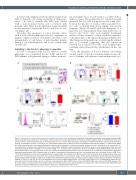
Functional switching of leukemic cells by stromal contact
A recent study implicated mitochondrial transfer from MSC to leukemic cells during acquisition of chemoresis- tance.27,28 In order to examine this, MSC were labeled with a mitochondrial tracker and co-cultured with leukemic cells. There was no difference in mitochondrial tracker intensity between the Sca-1(-) and Sca-1(+) sub- sets (Figure 1F).
Altogether, this emergence of a new leukemic subset with a stem cell-like phenotype (Sca-1(+)) represents an intrinsic cellular evolution of leukemic cells that occurs independently of cell fusion or mitochondrial transfer during in vivo leukemogenesis and in vitro culture with stromal cells.
Switching to the Sca-1(+) phenotype is reversible
In order to determine if the Sca-1(+) subset is a stable phenotype, we sort-purified Sca-1(+) (LSK) and Sca-1(-) (LK) leukemic cells generated during co-culture with stro-
ma, and replated for a second round of co-culture with or without stroma. The purified Sca-1(-) cell fraction again generated Sca-1(+) cells during the second round selec- tively in the presence of stroma, whereas purified Sca- 1(+) cells co-cultured with stroma rapidly decreased in frequency (Figure 2A and B) with the emergence of a major Sca-1(-) cell population. Thus, final stable ratios of Sca-1(+) and Sca-1(-) cells were similarly maintained under secondary stromal co-culture conditions regardless of the phenotype of the initial cell population (Figure 2C). The changes in cell populations occurred rapidly within 3 days of co-culture suggesting that the conversion between Sca-1(-) and Sca-1(+) cells occurs by phenotypic switching rather than selective proliferation in the cul- ture.
Thus, the emergence of Sca-1(+) leukemic cells during stromal contact occurs in a reversible manner in any sub- sets of leukemic cells without clonal predisposition (sto-
AB
CD
EF
Figure 1. Generation of a stem cell-like phenotype in a subset of leukemic cells. (A) Schematic illustration of the experiment. Murine acute myeloid leukemia (AML) cells were generated by transduction of fluorouracil (5-FU)-treated bone marrow (BM) cells with retrovirus encoding oncogene (MN1, or HoxA9/Meis1). Shown are retroviral vectors, experimental procedure for transplantation into mice, and the light microscopy morphology of transformed leukemic cells visualized by Giemsa staining. (B) Generation of Sca-1(+) (Lin-c-kit+sca-1+: LSK) leukemic cells during co-culture with murine mesenchymal stromal cells (mMSC). Co-cultures with mMSC for 3 days were performed in the presence (transwell) or absence (direct contact) of a transwell membrane between the cells in comparison to stroma-free (SF) cul- ture. Phenotypes of leukemic cells (CD45+GFP+) from co-cultured MSC (CD45-GFP-) were analyzed by flow cytometry. Shown are the representative profile (left) and quantification (right) (mean ± standard error of the mean [SEM] , n=7, *P<0.05). (C) In vivo generation of Sca-1(+) leukemic subsets. MN1 leukemic cells (Lin-c-kit+) were transplanted into mice and generation of Sca-1(+) subsets among BM engrafted leukemic cells were examined at 2 weeks post-transplantation (95% green fluorescent protein postive [GFP+] leukemic cells at the point). Representative flowcytometry plot (left) and quantification (right) are shown (mean ± SEM, n=10, *P<0.05). (D) Experimental scheme for analyzing cell fusion between MSC and leukemic cells. MSC transduced with a retroviral vector encoding yellow fluorescent protein (YFP), and leukemic cells transduced with a vector encoding GFP were co-cultured for 3 days. Shown are the experimental scheme (left) and representative flow cytometry profiles showing the absence of double positive (YFP/GFP) populations before and after co-culture (right). (E) Flow cytometry profiles for comparison between LK (Sca-1(-)), and LSK (Sca-1(+)) cell populations of cell size by forward scattering (left), and of DNA content (right) (n=5). (F) Experimental scheme to com- pare mitochondrial transfer between Sca-1(+) and Sca-1(-) cell populations. Murine MSC were pre-labeled with MitoTracker and co-cultured with MN1 leukemic cells for 3 days. Shown are representative flow cytometry plots from the experiments, each indicated leukemic cell subset (LK or LSK) of leukemic cells (CD45+GFP+) was gated and analyzed for MitoTracker and quantified for difference in mitochondrial transfer (n=4).
haematologica | 2022; 107(2)
383
