
Page 24 - 2022_02-Haematologica-web
P. 24
V. Krishnan et al.
the PRC2-EZH2 axis for survival and TKI resistance.73,74 Likewise, higher BMI1 levels at diagnosis correlated with disease progression from CP to BC.12 while BMI1 overex- pression in CP CD34+ cells increased proliferation and self-renewal,75 and transformed B-lymphoid progenitors in vivo.76
DNA methylation-associated gene expression changes
Many studies have examined the role of DNA methyla- tion as a regulator of aberrant GE in CML pathogenesis. In candidate-based approaches, genes involved in cell cycle regulation (P16, P53, PLCD1, PER3, HIC1), differentiation (HOXA4, DLX4, DDIT3, SPI1) proliferation (CDH13, DAPK1), apoptosis (BIM), Wnt regulation (sFRP1, CBY1), LSC maintenance (MTSS1), and cell signaling (Jun B, SOCS2) were identified as targets of DNA methylation.67,77
Recent unbiased genome-wide methylome analyses have solidified the concept of aberrant DNA methylation as a driver of resistance and transformation. The number of differentially methylated regions in CP increased from ~600 to ~6,500 CpG sites in BC.78 BC was associated with heightened DNA hypermethylation, and to a lesser extent hypomethylation, around promoters of genes involved in
stem cell fate, differentiation and leukemia-related func- tions.10 Mechanistically, differential DNA methylation pat- terns in CML have been attributed to underlying DNMT3A/TET2 mutations, PRC2-dependent epigenetic re-programming, and cytosolic sequestration of Tet2 by BCR-ABL1.79 Notably, the physiological targeting of DNA hypermethylation using 5-aza-2’-deoxycytidine amelio- rated disease phenotypes in a mouse model of CP dis- ease,80 while low-dose decitabine displayed clinical activi- ty in patients refractory to imatinib,81 suggesting DNA methylation does indeed contribute to TKI resistance.
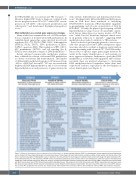
Based on the biological insights gleaned so far, it is pos- sible that progression-related DNA methylation signa- tures may already be evident at diagnosis, particularly in patients presenting with advanced CP.10 The DNA methy- lation status of specific target genes might therefore be useful in the timely identification of such patients for more aggressive therapies. Furthermore, given that DNA methylation is a relatively stable epigenetic and biochem- ical mark, there are practical advantages to developing DNA methylation-based biomarkers rather than tran- script-based readouts, especially for the development of robust clinical-grade tests (Figure 3).
Figure 3. Stages of development of gene expression-based biomarkers. In chronic myeloid leukemia (CML), the development of gene expression-based biomarkers can be divided into three stages following an initial discovery phase. These stages will each determine the analytical validity, clinical validity, and clinical utility of the tests in question. Examples of CML-specific issues or questions that are pertinent to each stage are outlned in boxes under each stage. GE: gene expression; IHC: immunohistochemistry; FC: flow cytometry; RT-PCR: reverse transcriptase polymerase chain reaction; ISH: in-situ hybridization; LCM: laser capture microdissection; scRNA-seq: single-cell RNA sequencing; ATAC-seq: assay for transposase-accessible chromatin sequencing; BM: bone marrow; PBMC: peripheral blood mononuclear cell; MNC: mononuclear cells; FFPE: formalin-fixed paraffin-embedded tissues; PB: peripheral blood; NK: natural killer cells; MDSC: myeloid-derived suppressor cells; TKI: tyrosine kinase inhibitor; NCCN: National Comprehensive Cancer Network; ELN: European LeukemiaNet; BC: blast crisis; EFS: event-free survival; DFS: disease- free survival; PFS: progression-free survival; OS: overall survival, TFR: treatment-free remission; 95% CI: 95% confidence interval; DMR: deep molecular response..
366
haematologica | 2022; 107(2)
