
Page 193 - 2022_02-Haematologica-web
P. 193
Sulfated non-anticoagulant heparin in SCD treatment
AB
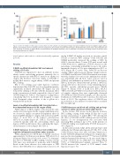
Figure 2. Effect of S-NACH on HbS oxygen binding affinity. (A) The sulfated non-anticoagulant heparin derivative (S-NACH) increases hemoglobin oxygen affinity. Aliquots of hemolysates from the sickling assay were subjected to p50 analyses using the Hemox Analyzer. (B) Representative curves show a dose-dependent left shift, indicating an increase in oxygen affinity. Summarized data for biological replicates (n=5) are indicated in the graph. The findings confirm the primary direct anti-sickling mechanism of S-NACH.
tistical analyses, and results are considered statistically significant if P<0.05.
Results
S-NACH modified intracellular HbS and reduced sickling of SS cells
S-NACH was engineered to have an aldehyde moiety, which confers anti-sickling properties primarily due to specific interactions with HbS to increase its affinity for oxygen. We therefore tested the ability of S-NACH to modify HbS, increase oxygen affinity of HbS, and prevent RBC sickling.
Our in vitro sickling assay under hypoxic conditions demonstrated that S-NACH, in a dose-dependent manner, significantly modified intracellular Hb (Figure 1A) and reduced the sickling of SS cells, with the maximum effect at the concentration of 2 mM, comparable to that of 1 mM GBT440 (Figure 1B; Table 1). This supports our hypothesis considering that two molecules were designed to target both N-terminal valine residues of the a globin in a tetrameric Hb molecule.
Levels of modified intracellular HbS translated into a dose-dependent increase in Hb oxygen affinity
When aliquots of HbS-complex solution from the same studies were investigated in the oxygen equilibrium assay, we observed a similar concentration-dependent effect on increasing HbS affinity for oxygen, with a reduction in P50 values of about 55% at the highest concentration (65.7±3.2 at 2 mM) (Figure 2A; Table 1). These findings correlated linearly with the anti-sickling effects and degrees of HbS modifications, thus confirming this target- ed mechanism of action (Figure 2B; Table 1).
S-NACH decreases in vivo red blood cell sickling and regulates inflammatory cytokines under normoxia
When administered to C57/B mice, S-NACH caused an approximately 3-fold increase in plasma TFPI after 2 h of treatment (Figure 3A) at both doses tested. To determine the effect of S-NACH on RBC sickling, total blood from SCD mice was incubated at normoxia with S-NACH. Based on the lower effective dose with respect to TFPI release, the S-NACH dose for animal studies was set at 10
mg/kg. 5-HMF (10 mg/kg) was used as a positive control because it decreases RBC sickling.33 Both S-NACH and 5-HMF moderately decreased the sickling of RBC by 35-50% (data not shown). Townes SCD mice treated with S-NACH showed a significant (*P<0.05) decrease in the percentage of circulating sickled RBC for up to 4 h, with a maximum decrease at 2 h after administration (50% to 35%) (Figure 3B, C) (n=6). Thus, S-NACH can decrease sickling of RBC under normoxia. Plasma samples (untreat- ed, 5-HMF, 2 and 6 h after S-NACH treatment) were quan- titatively analyzed for various pro-inflammatory media- tors (interleukin [IL] 1b, IL-6, tumor necrosis factor-a [TNF-a]), anti-inflammatory mediators (IL-10, interferon g [IFN-g], monocyte chemoattractant protein 1 [MCP-1]), and growth factors (monocyte colony-stimulating factor [M-CSF], vascular endothelial growth factor [VEGF]) (Figure 4). Plasma levels of IL-1b, IL-6, IFN-g, MCP-1, TNF-a, M-CSF, and VEGF were increased in SCD untreat- ed samples, whereas they were significantly decreased (P<0.0005) in S-NACH-treated samples, at both 2 and 6 h. Furthermore, S-NACH was able to increase the decreased levels of 1L-10. The regulatory effect of S-NACH was most effective at 2 h, similar to its effectiveness on sickled RBC morphology.
S-NACH decreases red blood cell sickling and prolongs survival of sickle cell disease mice under hypoxia
In the Townes SCD mouse model, hypoxia increases RBC sickling, causing death within 15 min due to pul- monary sequestration of sickled RBC.26 We investigated the effect of S-NACH on RBC sickling and survival under hypoxia. Ex vivo deoxygenation was associated with increasing RBC sickling of up to 70%. In the presence of S-NACH, sickling was significantly (P<0.05) decreased to 30% (Figure 5).
In the survival study, while all the untreated mice died within the first 15 min under hypoxia (5% O2), 50% of the S-NACH-treated mice were alive at 30 min, which was 1 h after S-NACH administration (Figure 6), a typical times- pan used for investigating survival.33,40,41 Because S-NACH exhibited maximal effectiveness at 2 h, mice under hypox- ia were observed for an additional 1 h, during which 37.5% of S-NACH-treated animals survived. Thus, S- NACH increased the survival of SCD animals under hypoxia for up to 3 h.
haematologica | 2022; 107(2)
535
