
Page 89 - 2022_01-Haematologica-web
P. 89
Mechanisms of sorafenib resistance in AML
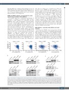
phospho-ERK were evident in TSC1-deficient, but not in TSC2-deficient cells, potentially reflecting different roles of TSC1 and TSC2 in the TSC complex. Levels of RAS protein were elevated in LZTR1-deficient cells, but not in those deficient in NF1 or TSC1 (Figure 2C).
MTOR and MAPK pathways are upregulated in acute myeloid cells resistant to FLT3 inhibitors
In a parallel approach to understand mechanisms of resistance to sorafenib, we generated AML cell lines resistant to FLT3 inhibitors by gradually exposing MOLM13 cells to type I (crenolanib) or type II (quizar- tinib and sorafenib) FLT3 inhibitors. Crenolanib- and sorafenib- resistant MOLM13 cells showed reduced sen- sitivity, detected by higher IC50 and AUC values, to both type I and type II inhibitors, whereas quizartinib-resis- tant cells showed resistance only to type II inhibitors (Figure 3A). To investigate whether there is an overlap between the acquisition of resistance by CRISPR-derived knockout cells versus resistance generated by prolonged exposure to drugs, we evaluated the activity of MTORC1 and MAPK pathways. We detected an increase in levels of phospho-ERK and elevated RAS lev- els in FLT3 drug-resistant cells, indicating upregulation of the MAPK pathway (Figure 3B). Surprisingly, levels of TSC2 were increased in crenolanib- and sorafenib-resis-
A
B
tant cells; in contrast, loss of function for TSC2 was revealed in the CRISPR knockout resistance screen. Previous studies showed that the MAPK pathway can inhibit MTOR activity by phosphorylating TSC2 at S664 causing dissociation of the TSC complex observed in breast and colon carcinomas.41-43 This observation prompted us to test for levels of phospho-TSC2. We observed enhanced levels of phospho-TSC2 at S664 in these two cell lines, suggesting that inhibition of TSC complex formation by the MAPK pathway promotes resistance to FLT3 inhibitors. Moreover, we observed an elevated level of phospho-TSC2 at Y1571, which addi- tionally impairs the TSC1-TSC2 interaction,44 supporting inactivation of TSC2 in these resistant cells, concordant with our CRISPR screen results.
MEK inhibitors resensitize FLT3-inhibitor-resistant cell lines
Data from the CRISPR screen and resistant cell lines suggested that upregulation of the MAPK signaling path- way contributes to resistance to FLT3 inhibitors in AML. This prompted us to hypothesize that inhibitors target- ing the MAPK pathway may resensitize FLT3-inhibitor- resistant cells. Parental and resistant cells were tested for sensitivity to the MEK inhibitor trametinib and MTOR inhibitors PP242, PI-103 and rapamycin. Resistant cells
Figure 2. Low sensitivity to sorafenib correlates with low expression levels of CRISPR top hits LZTR1, NF1, and TSC2 in specimens from acute myeloid leukemia patients and inactivation in the MOLM13 acute myeloid leukemia cell line reveals aberrant activation of MAPK or MTOR pathways. (A) Scatter plots of RNA expres- sion levels (shown as normalized reads per kilobase per million [RPKM]) plotted against drug sensitivity measured ex vivo as area under the curve (AUC) from samples from patients with acute myeloid leukemia harboring FLT3-ITD mutations (n=86). Pearson correlation coefficients are shown as (r) values Statistical significance is indicated. ****P<0.0001, ***P<0.001, **P<0.01, n.s., non-significant. (B, C) Immunoblot analyses of proteins from LZTR1-, NF1-, TSC1- and TSC2-deficient cells (LZTR1_sgRNA, NF1_sgRNA, TSC1_sgRNA and TSC2_sgRNA, correspondingly) in comparison to parental and non-targeting (NT_sgRNA) controls with indicated anti- bodies. p-ERK denotes phosphorylated-ERK (Thr202/Tyr204), p-MTOR denotes phosphorylated-MTOR at S2448. Vinculin and GAPDH immunoreactivity served as loading controls.
C
haematologica | 2022; 107(1)
81
