
Page 82 - 2022_01-Haematologica-web
P. 82
B.Z. Carter et al.
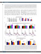
Figure 7. Protein levels in various leukemia cell populations in mouse bone marrow as determined by CyTOF. Bone marrow (BM) cells were collected from the mice on day (d) 25 of treatment (A to C) or from moribund mice (D to F) from each treatment group (n=2 or 3 mice/group). (A and D). BCL-2, MCL-1, and c-MYC levels in various leukemia cell populations in each treatment group. (B and E) Cell surface expression levels of CXCR4 and CD44 (B) or CXCR4 (E) in various cell populations in each treatment group. (C) β-CATENIN and p-AKT levels in CD45+ and CD34+CD38-CD123+ cells. (F) p-AKT levels in various cell populations. The protein levels of individual samples are presented as heat maps. After stained with antibodies against cell surface markers, all BM samples collected at d 25 treatment were bar- coded, pooled into the same tube, stained, and run concomitantly and all BM samples collected from moribund mice were barcoded, pooled into the same tube, stained, and run concomitantly.
tiveness of the venetoclax plus azacitidine combination in its ability to suppress OxPhos, disrupt the TCA cycle, and perturb cell energy metabolism, thereby efficiently target- ing leukemia stem cells, an effect that was not achieved with the venetoclax treatment alone.7 We found that MCL-1 regulates redox and metabolic functions in AML cells and that genetic or pharmacological inhibition of MCL-1 suppressed several cellular energetic and metabolic
A
B
pathways, including TCA cycle, glycolysis, and PPP. We further demonstrated that inhibiting this function of MCL-1 contributed to the enhanced activity of venetoclax against AML cells.
ROS stabilize HIF1 thereby activating hypoxia signal- ing.45,46 Although MCL-1 alteration in AML cells did not elic- it detectable changes in HIF1a levels, it elicited changes in CXCR4, a HIF1a target, and CD44. Both, the
C
Figure 8. Effects of BAX and/or BAK knockdown on apoptosis, mitochondrial respiration, and migration/adhesion to mesenchymal stromal cell of acute myeloid leukemia cells in responses to MCL-1 inhibition. OCI-AML3 cells were transfected by electroporation with control, Bax, Bak, or Bax and Bak small interfering RNA (siRNA) (4 mM) for 48 hours (h) and then treated with AZD5991 (50 nM) for 24 h. (A) BAX and BAK levels in siRNA-transfected OCI-AML3 cells at 48 h, determined by western blot. The left panel is the result of a representative experiment and the right is the quantitative results of three independent experiments. (B) Apoptosis by flow cytometry and mitochondrial respiration by Seahorse of siRNA-transfected OCI-AML3 cells treated with AZD5991 at various time points. (C) Apoptosis and migration (6 h) and adhesion (24 h) of siRNA-transfected OCI-AML3 cells treated with AZD5991 for 24 h compared with untreated controls. The experiments were performed in triplicates (with duplicates for each experiment in the Seahorse experiment). The results are expressed as mean ± standard error of the mean. AnnV: annexin V; OCR: oxygen consumption rate. MSC: mesenchymal stromal cells.
74
haematologica | 2022; 107(1)
