
Page 70 - 2022_01-Haematologica-web
P. 70
B.Z. Carter et al.
G
H
I
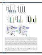
J
Figure 1. MCL-1 regulates cellular metabolic functions, and genetic or pharmacological inhibition of MCL-1 decreases mitochondrial respiration and metabolism in acute myeloid leukemia cells. (Ai) Seahorse analysis of mitochondrial respiration in MCL-1-overexpressing (MCL-1-OE), MCL-1 knockdown (MCL-1-KD), and (Aii) AZD5991-treated (24 h) OCI-AML3 cells. For MCL-1-OE and –KD cells, bulk populations were used unless indicated otherwise. (B) Cellular reactive oxygen species (CellROS) and mitochondrial ROS (MitoSOX) in MCL-1-OE and MCL-1-KD OCI-AML3 cells. (C) Glutathione (GSH) and GSH/glutathione and glutathione disulfide (GSSG) ratios in MCL-1-OE and MCL-1-KD OCI-AML3 cells. (D to I) OCI-AML3 MCL-1-KD clone (shC16) with an approximately 65% MCL-1 reduction (Online Supplementary Figure S1D) and OCI-AML3 cells treated with 50 nM AZD5991 were used for metabolomics experiments. WT: wild-type untreated control. (D) Relative enrichment of 13C from 13C -1,2-glucose or 13C -glutamine (M>0) into key tricarboxylic acid (TCA) cycle intermediates. (E) Fractional enrichment of 13C from 13C -glutamine for citrate,
2
fumarate, and malate. (F) Relative lactate levels in medium after 6 hours (h) of incubation with new medium. Lactate levels were determined by summing MS1 ion intensities for all isotopes in the 13C -1,2-glucose (left) and the 13C -glutamine tracer experiment (right). (G) Fractional enrichment in extracellular lactate after 6 h of
5
5
5
5
2
incubation in the 13C -1,2-glucose (left) and 13C -glutamine (right) tracer experiments. (H and I) Relative pentose phosphate pathway (oxPPP) flux (left) and 6-phospho-
2
gluconic acid (6PG) levels (right, H) and relative levels of unlabeled ATP and 13C enrichment into ATP (I). Average WT and shVec M+0 levels were set to a relative level of 1. WT: parental OCI-AML3 cells; AZD5991: AZD5991-treated OCI-AML3 cells; shVec: vector control OCI-AML3 cells; shC16: MCL-1-KD OCI-AML3 cells. *P≤0.05, **P≤0.01, ***P≤0.001, ****P≤0.0001. Experiments were performed in triplicates (for seahorse analysis: duplicates for each experiment). (J) Schematic illustration of carbohydrate metabolic pathways decreased by MCL-1 inhibition. Cit: citrate; Fum: fumarate; Mal: malate; aKG: a-ketoglutarate; Oaa: oxaloacetic acid; MFI: mean fluorescence intensity.
OCI-AML3 migration toward, and adhesion to, MSC (Figure 2B). Consistently, inhibition of MCL-1 with AZD5991 decreased OCI-AML3 surface CXCR4 and CD44 expression and interaction with MSC (Figure 2C). Like other MCL-1 inhibitors, AZD5991 increased levels of MCL-1 (Figure 2C). BCL-2 inhibition with venetoclax in
OCI-AML3 cells did not appreciably decrease CD44 sur- face expression, AML cell migration, or adhesion to MSC. However, it decreased CXCR4 surface expression (Figure 2D). These results suggest that MCL-1 enhances surface adhesion molecule expression and promotes leukemia- stroma interactions.
62
haematologica | 2022; 107(1)
