
Page 357 - 2022_01-Haematologica-web
P. 357
Case Report
ily and past medical history of malignancy may be informative, as ALPS-FAS and other germline diseases are known to predispose to lymphoma and solid tumors. However, in cases of adult-onset ALPS-FAS, the diagno- sis may still be challenging as other hematologic diseases may have overlapping clinical and pathologic features, including angioimmunoblastic T-cell lymphoma, Rosai- Dorfman disease, iMCD, and IgG4-RD. Rarely, ALPS may co-occur with one of these disease entities and as such mask the typical morphologic features of ALPS.5,6 While certain disease working groups, including that for iMCD, currently recommend evaluation for ALPS, the current consensus diagnostic criteria for IgG4-RD do not.7 In our reported case, if flow cytometry had not been performed to further investigate the increased αβ+DNT, the diagnosis of ALPS would likely have been missed as the lymph node histopathological features
were masked by IgG4-RD. Thus, as previously suggested by van de Ven and colleagues,6 screening for increased αβ+DNT by flow cytometry or ALPS-associated muta- tions by NGS should be considered in patients with IgG4-RD, particularly in those with other clinical, patho- logic, or laboratory features characteristic of ALPS. Identification of ALPS-FAS patients with concurrent IgG4-RD may have significant therapeutic implications, as patients may require chronic therapy or become intol- erant to standard immunosuppressive therapy, and thus may benefit from targeted, steroid-sparing therapy such as rituximab or sirolimus.8
A mechanistic link between the pathogenesis of ALPS and IgG4-RD is currently unknown. B lymphocytes are involved in the pathogenesis of IgG4-RD as evidenced by marked clinical responses to B-cell-directed therapy with rituximab. It is hypothesized that the oligoclonal
A
E
BF
CG
DH
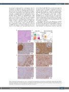
Figure 3. Expansion of double-negative αβ+ T cells in a background of immunoglobulin G4-related disease. (A) Right level V cervical lymph node excision stained with hemoxylin and eosin (H&E), (B) CD20, (C) immunoglobulin G4 (IgG4) and (D) IgG. (E, left) Flow cytometry analysis of lymph node gated on CD3+ lymphocytes showing CD4 vs. CD8 and (E, right) gated on CD3+CD4-CD8- showing TCRαβ vs. TCR𝛾𝛿. (F) Immunostaining for CD3, (G) CD4 and (H) CD8. For all micrographs, a 20x objective was used and 200x total magnification is presented.
haematologica | 2022; 107(1)
349
