
Page 356 - 2022_01-Haematologica-web
P. 356
Case Report
AA
B
C
D
B
C
D
Figure 1. Increased immunoglobulin G4 postive plasma cells in a lymph node with multicentric Castleman disease-like features. Right cervical lymph node excision stained with hemoxylin and eosin (H&E). (A) 2x objective, 20x total magnification and (B) 20x objective, 200x total magnification). (C) Immunoglobulin G4 (IgG4) 20x objective, 200x total magnification and (D) IgG 20x objective, 200x total magnification.
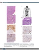
Figure 2. Radiographic and pathologic features of immunoglobulin G4-relat- ed disease. (A) Positron emission tomography maximum intensity projection image showing hypermetabolic lymphadenopathy above and below the diaphragm along with lesions in the pancreas and spleen . (B) Pancreatic core needle biopsy stained with hemoxylin and eosin (H&E), (C) immunoglobulin G4 (IgG4) and (D) IgG. For all micrographs, a 20x objective was used and 200x total magnification is presented.
348
haematologica | 2022; 107(1)
