
Page 346 - 2022_01-Haematologica-web
P. 346
Letters to the Editor
A novel index to evaluate ineffective erythropoiesis in hematological diseases offers insights into sickle cell disease
Ineffective erythropoiesis (IE) is a significant patholog- ical factor in many types of anemia including b-thalassemia and myelodysplasia and has more recently been described in sickle cell disease (SCD). We have eval- uated a novel index of ineffective erythropoiesis (IoIE) as a way of quantitating IE and facilitating its comparison between patients, conditions, and the effects of treat- ments. We calculated IoIE by dividing the plasma con- centration of soluble transferrin receptor (sTfR, nmol/L) (proportionate to the volume of erythroid tissue) by the absolute reticulocyte count (ARC, x109/L) (effective ery- throid output from the bone marrow). The upper limit of normal IoIE was calculated at 0.28, using the established normal ranges of sTfR and ARC, and confirmed using control samples. We studied 414 SCD patients and show that IE is a feature of patients with HbSS (median IoIE 0.37), but not HbSC (median IoIE 0.27). We validated IoIE as a measure of IE in a cohort of 44 patients with HbE-beta thalassemia, a condition in which IE is known to play a major part and, as expected, found high levels (median IoIE 1.46). Within the HbSS cohort, we find higher HbF levels associate with reduced IE and that transfusion reduces IE, whereas hydroxyurea (HU) treat- ment appears to lead to increased IE. This index is a sim- ple and meaningful measure of IE, showing that it is clin- ically important in SCD, and suggesting novel therapeu- tic approaches.
IE is the abnormal differentiation of erythroid progeni- tors, with an expanded progenitor compartment,
increased erythroblast destruction and a relative paucity of reticulocytes produced compared to the volume of the erythron.1 IE is well described in beta-thalassemia but less so in SCD, where anemia is generally attributed to hemolysis.2 There is, however, evidence of dysfunctional erythroid differentiation in SCD. Erythroblasts isolated from the bone marrow of SCD patients sickle under hypoxic conditions3 whilst analysis of chimeric hematopoiesis in non-myeloablative transplanted SCD patients have demonstrated a survival advantage of the donor erythroid progenitor cells.4 More recently, high levels of apoptosis between the polychromatic and orthochromatic stages were identified, with fetal hemo- globin (HbF) being a key protective factor against this.5
Quantitation of IE in patients would facilitate its study in SCD and other anemias and allow an assessment of novel treatments. Transferrin receptor (CD71) is a mem- brane protein expressed during erythropoiesis. It is also released into the circulation, with its serum concentra- tion shown to be proportional to the mass of erythropoi- etic tissue.6,7 Soluble transferrin receptor (sTfR) levels are elevated in SCD, reflecting an increased erythropoietic drive, but show no correlation with disease severity.8 We propose a meaningful representation of IE that can be determined by measuring the ratio of the mass of ery- thropoietic tissue (sTfR) to the erythroid output from the marrow (ARC), analogous to the way the reticulocyte percent correlates with the rate of hemolysis.9 Where IE is occurring, the erythron mass will be large, with higher sTfR levels, whilst the relative output of reticulocytes (ARC) will be lower, and the ratio will be higher. We refer to this ratio as the Index of Ineffective Erythropoiesis (IoIE), and explore its significance in patients with SCD.
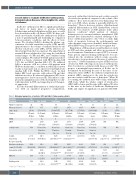
Table 1. Biological parameters of controls, SCD and HbE/b-thalassaemia patients.
Genotype
Treatment
Number of patients Mean age in years (min - max) Gender
G6PD gHbF
a-thalassemic
AA
Control
22 42.77 (16-93) 8 Female, 14 Male
SS
Transfusion
58 29.17 (16.94-51.75) 26 Female, 32 Male
SC
Non- treated
59 Female, 8 Female, 11 Female, 7 Female, 28 Male 4 male 12 Male 15 Male
Non- treated
182
31.7 (17.3-63.58) 110 Female, 72 Male
HC
50 28.71 (18.1-57.46) 28 Female, 22 Male
HbE/b-thalassemia Non- treated Transfusion
23 22
11 18.5 (17.9-74.9) (18.93-68.28) (1-39) (4-53)
S/b + Non- treated
87 39.15
12 42.48
0 11 4 0 2 1 0 0
N/A 2.718 2.135 2.137 (1.89-2.62) (1.89-2.69) (1.89-2.49)
2.1777 2.144 (1.89-2.49) (1.89-2.32) 62 aa /aa, 8 aa /aa, 23aa/a-, 3aa/-a,
2 a-/a (23.1-99.6)
208.1
115.4 (80-147.5) 1.836 (0.2-11.1) 41.34 (14-143)
N/A N/A N/A N/A
N/A
131 aa /aa, 39 aa /aa, 31 aa /aa, 38aa/a-, 17aa/a-, 15aa/a-,
20.66 (10.6-23.8)
289.2 132.6 (121.5-600.3) (18.8-404.8)****
112.8
N/A 84.16 (42-124.3)
N/A 6.8 (0.2-25.6)
N/A 104.8 (10.6-800)
13a-/a- 123.9 (41.4-241.1)
2a-/-a 82.95 (27.8-187.5)****
4a-/a- 103.9 (33.26-202)****
1a-/a- 51.72 (27.93-90.38)
Soluble
Transferrin
Receptor (nmol/L)
Absolute reticulocyte
count(100*10x9/L) (48.6-214.4) (152.6-695) (148.4-888.9)**** (140.3-684.8)**** (91.53-442.1) (67.97-350.8) (100.6-1320) (3.4-267.9)****
366.6
384.5
95.96 (61.8-121)**** 3.03 (0.2-11.1)**** 59.45 (21.3-187)****
286.5
88.44 (41.6-113.8)* 10.38 (1.25-29.02)**** 112.3 (24.3-354.3)
174.4 122.1
5.3 (0.7-11.6) 29.84 (9.6-81.15)
262 90.37 71.57 94.5
Hemoglobin (g/dL) HbF (%) Erythropoietin
(10-105)
45.36 35
(11.9-132)*** N/A N/A
(8.9-66)
***P<0.001 ****P<0.0001.
338
haematologica | 2022; 107(1)
