
Page 336 - 2022_01-Haematologica-web
P. 336
Letters to the Editor
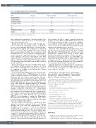
Table 1. Serological characteristics of the patients Healthy donor
Patient 1
Positive (1,680 AU) 10.4
0
2.9
3.2
1.8
Positive Positive
Patient 2
Positive (1,290 AU) 0
0
0
0.7
1.4
Positive Positive
aPF4
aCL IgG (GPL/mL) aCL IgM (MPL/mL)
a 2-GPI IgG (UA/mL)
a 2-GPI IgM (UA/mL) LA
aCL
(TLC Immunostaining)
aVim/CL
Negative 0
0
0
0 0.95
Negative Negative
aPF4: anti-platelet factor 4 antibodies; aCL: anti-cardiolipin antibodies; a 2-GPI: anti- 2 glycoprotein I antibodies; LA: Lupus anticoagulant; TLC: thin-layer chromatography; aVim/CL: anti-Vimentin/Cardiolipin complex antibodies.
three independent experiments. Statistical analysis was performed by the paired Student’s t-test. Statistical signif- icance was set up at P≤0.01.
Results show that the treatment with both IgG frac- tions from patients with VITT, induced a significant increase of both phospho-ERK (Figure 1A) and phospho- p38 expression (Figure 1B) compared to untreated platelets or treated with healthy donor IgG obtained from a representative healthy donor vaccinated with ChAdOx1 nCoV-19. As a consequence of ERK activation, we also observed a significant increase of TF levels in treated platelets. Moreover, pretreatment with ERK inhibitor PD98059 partially prevented TF expression in samples stimulated with patient IgG fractions (Figure 1C), indicating a functional link between ERK activation and TF expression.
Surface TF expression was also verified by flow cytom- etry analysis, which revealed an increase of TF in platelets treated with IgG fractions from the two patients as compared to healthy donor IgG fraction (Online Supplementary Figure S1B).
Sera of patients and healthy donor were tested for anti- bodies to platelet factor 4 (PF4)/polyanion by using a commercial enzyme immunoassay (Immucor, Lifecodes, Waukesha, WI). Sera with an optical density >500 arbi- trary units (AU) were considered as positive.3 In addition, anticardiolipin (aCL, IgG, IgM) and anti-b2-glycoprotein I (b2-GPI, IgG, IgM) were detected by chemiluminescence assay, using Zenit RA Immunoanalyzer (A. Menarini Diagnostics, Florence, Italy). Finally, aCL were also detected by TLC immunostaining and anti-vimentin/car- diolipin (aVim/CL) antibodies by ezyme-linked immunosorbant assay, as previously described;10 lupus anticoagulant (LA) was analyzed by two coagulation sys- tems, a dilute sensitized activated partial thromboplastin time (aPTT) and a dilute Russell’s viper venom time (dRVVT), also performing a confirm test (Hemoliance Instrumentation Laboratory, Lexington, MA, USA). Serological characteristics of the two patients and healthy donor are summarized in Table 1.
Previous studies described that a pathogenic PF4- dependent syndrome, unrelated to the use of heparin therapy, can occur after the administration of the ChAdOx1 nCoV-19 vaccine.2,3,4-6 All subjects displayed anti-PF4 antibodies which were able to activate platelets obtained from healthy donors.2 A subset of these anti- bodies may activate platelets after binding to PF4/heparin complexes, causing the prothrombotic adverse drug reac- tion HIT. In autoimmune-HIT, anti-PF4/P-antibodies acti- vate platelets in the absence of heparin. In the present study we observed for the first time that IgG fractions of
these patients are able to trigger a signal transduction pathway involving ERK and p38 MAPK, which may lead to TF expression increase with a consequent amplifica- tion of platelet activation and coagulation cascade.11 We cannot exclude the possibility that other additional signal transduction pathways may be involved.
However, other autoantibody specificities have been described in sera of patients affected by VITT, including aPL and/or LA.2,4 This finding may be relevant, since anti- b2GPI/b2GPI complexes were shown to induce signal transduction pathway(s) leading to platelet activation,12 particularly promoting thrombosis via p38 MAPK13 and consequent involvement of TF as major initiator of the clotting cascade. We confirmed and extended the analy- sis of aPL, also showing the presence of “unconventional” (“non-criteria”) aPL in serum of our patients.14
How the pathogenic mechanism may be consequent to ChAdOx1 nCov-19 vaccination remains unclear;15 how- ever, a possible pathogenic role of the adenoviral viral vector cannot be excluded.
Roberta Misasi,1 Antonella Capozzi,1 Gloria Riitano,1 Serena Recalchi,1 Valeria Manganelli,1 Vincenzo Mattei,2 Agostina Longo,1 Manuela De Michele,3 Tina Garofalo,1 Fabio M. Pulcinelli1 and Maurizio Sorice1
1Department of Experimental Medicine, "Sapienza" University of Rome, Rome; 2Biomedicine and Advanced Technologies Rieti Center, Sabina Universitas, Rieti; 3Emergency Department, "Sapienza" University of Rome, Rome, Italy
Correspondence:
MAURIZIO SORICE- maurizio.sorice@uniroma1.it doi:10.3324/haematol.2021.279729
Received: August 3, 2021.
Accepted: September 29, 2021.
Pre-published: October 7, 2021.
Disclosures: no conflicts of interest to disclose.
Contributions:FMP and AC designed and performed the research; MDM selected the patients; GR, SR and VM performed experiments; AL and VM provided and analyzed the data; RM and TG wrote the paper; MS designed, organized, and supervised the research and edited the paper. All authors read, edited, partici- pated in the revision, and approved the manuscript.
References
1. Hu B, Guo H, Zhou P, Shi ZL. Characteristics of SARS-CoV-2 and COVID-19. Nat Rev Microbiol. 2021;19(3):141-154.
2. Scully M, Singh D, Lown R, et al. Pathologic antibodies to platelet
328
haematologica | 2022; 107(1)
