
Page 322 - 2022_01-Haematologica-web
P. 322
Letters to the Editor
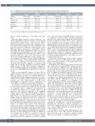
Table 1. Peripheral blood cell counts in in utero thirdhand smoke- and clean air–exposed male and female mice.
Cell type
Platelets
MPV
Red blood cells Lymphocytes Monocytes Granulocytes HCT
Clean air
627.40 ± 88.57 4.84 ± 0.27 6.92 ± 0.50 1.40 ± 0.40 0.13 ± 0.06 1.86 ± 0.52 32.00 ± 2.25
Male
in utero THS 649.20 ± 42.13
4.82 ± 0.08 7.45 ± 0.27 1.93 ± 0.70 0.09 ± 0.02 2.39 ± 0.80
34.13 ± 1.41
P value 0.63
0.89 0.11 0.18 0.12 0.24 0.16
Clean air
504.80 ± 46.98 4.82 ± 0.13 6.71 ± 0.83 1.60 ± 0.49 0.13 ± 0.06 1.86 ± 0.50 31.98 ± 4.05
Female
in utero THS 517.30 ± 7 7.80
4.86 ± 0.11 6.69 ± 0.57 2.17 ± 0.59 0.09 ± 0.02 2.51 ± 0.67 31.06 ± 3.17
P value 0.77
0.62 0.96 0.13 0.12 0.14 0.69
All counts are expressed as thousands per microliter, except for red blood cells, which are expressed as millions per microliter. Data are presented as mean ± standard deviation.HCT: hematocrit;MPV:meanplateletvolume;THS:thirdhandsmoke.
reveal a statistical significance, with either of the ago- nists.
Given that platelet granule secretion is known to con- tribute significantly to platelet activity,8 we investigated agonist-induced ATP release and P-selectin surface expression as markers for dense and a-granules release, respectively. Dense granules as well as a-granules secre- tion were increased in platelets obtained from in utero THS exposed mice, in response to either ADP or throm- bin (Figure 2B; Online Supplementary Figure S1i and ii). These data revealed that platelet secretion contributes to the THS prothrombotic phenotype. In terms of sex- dependent differences, the in utero THS-exposed males showed much higher ADP-induced dense granule secre- tion compared to females, but no statistical difference was observed in the clean air-exposed mice (Figure 2B). Moreover, no differences between the two sexes were observed with thrombin regardless of exposure type (Figure 2B). In contrast, a-granules secretion was signifi- cantly elevated in THS exposed females compared to males following stimulation by thrombin; but this was not the case with ADP (Online Supplementary Figure S1i and ii).
Next, we investigated the impact of in utero THS on aIIbb3 activation; which was more pronounced in the THS exposed mice, in response to 5 μmol/L ADP and 0.1 U/mL thrombin (Online Supplementary Figure S1iii and iv); which is in accordance with the enhanced aggregation response and was demonstrated in both sexes. Interestingly, our analysis revealed a significant sex- based difference (higher in males) with both agonists.
As platelets are activated, phosphatidylserine (PS) becomes exposed at their outer surface, for the assembly of coagulation factor complexes.9 Subsequently, we determined the impact of in utero THS exposure on PS expression. We found PS expression to be markedly enhanced upon stimulation with thrombin or ADP fol- lowing THS in utero exposure (Online Supplementary Figure S1v and vi), which was documented in males and females. However, when both sexes were compared, it was found that the ADP effects were more pronounced in THS-exposed females compared to males. In contrast, when thrombin was analyzed, the effects in males were found to be higher than females. This discrepancy between the release of dense granules versus a-granules in males and females might be attributed to the fact that the former was performed using platelet-rich plasma whereas the latter using washed platelets; given that the presence of other plasma factors makes it difficult to assess whether the sex difference is inherent to platelets or related to plasma.10 As for PS exposure, female platelets showed more significant elevation in compari-
son to those from males with ADP. However, this trend is completely reversed when thrombin-stimulated platelets were utilized with a significant elevation in PS in males compared to females. This could be explained by the variation in dose-response between males and female platelets.11 It also should be noted that these are different platelet functional responses. In addition, anal- ogy could be inferred from the race disparity of thrombin PAR4 receptor that triggers enhanced platelet aggrega- tion as well as calcium mobilization when activated in African lineage compared to Caucasian.12 Similarly, a sex disparity in the receptors of different agonists or their downstream signaling pathways could explain the afore- mentioned discrepancies.
Collectively, our functional assays provide evidence that in utero exposure to THS triggers a state of platelet hyperactivity and contributes to the prothrombotic phe- notype in the offspring mice.
In summary, these data provide evidence that the neg- ative health effects of maternal THS exposure extend to the “non-exposed” offspring. Thus, our findings docu- ment for the first time that in utero THS exposure drives platelets into a state of hyperactivity, that manifests in a host of enhanced functional responses (e.g., aggrega- tion). Together, these effects ultimately lead to a pro- thrombotic phenotype. It is noteworthy that this danger, according to our current and published data, does not only affect “directly” THS-exposed mice as we have shown before6 but expands to the offspring of the exposed pregnant mice as well. Interestingly and impor- tantly, this prothrombotic phenotype endured despite the fact that offspring mice were not exposed to THS as they were moved to clean-air exposed cages until they reached 8-10 weeks of age. These data also highlight the underestimated risk of exposure to THS toxicants that persist up to several months after the last smoking has taken place.1,13 It is also important to note that this phe- notype is consistent with the state of hyperactive platelets we reported previously as a result of exposure to other forms of tobacco that are perceived as safe, namely e-cigarettes14 and hookah/waterpipe.15
As for the comparisons between males and females, although no sex differences could be demonstrated in bleeding time, thrombosis or platelet aggregation, we did observe significant differences in dense and a-gran- ule secretion, aIIbb3 activation as well as PS exposure when compared sex-wise.
In conclusion, our data clearly demonstrates for the first time that in utero THS exposure modulates the platelet biology in the non-exposed offspring, making them more susceptible to cardiovascular diseases.
314
haematologica | 2022; 107(1)
