
Page 297 - 2022_01-Haematologica-web
P. 297
Letters to the Editor
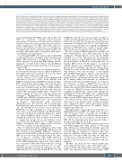
Figure 2. Maraviroc enhanced DNA damage induced by trabectedin. Hodgkin and Reed-Sternberg (HRS) cells were treated with maraviroc (100 mM), trabecte- din (500 pM) and their combination for 24 h. (A) Western blot for g-H2AX and a-tubulin protein expression in cHL cell lines. Membrane strips were incubated with mouse anti-phospho-histone H2A.X (Ser139) (clone JBW301) (Millipore) and mouse anti-a-tubulin antibody clone B-5-1-2 (Sigma Aldrich) and revealed with donkey anti-mouse IgG (H+L chain) A90-137P (Bethyl Laboratories). Images were acquired using a ChemiDoc XRS system (Bio-Rad). Data are representative of four experiments. (B) Bar charts showing densitometric analysis of g-H2AX expression normalized to a-tubulin as a loading control. Protein quantification was performed using ImageJ software. Values are means and standard deviation of four experiments. *P<0.05, treatments vs. control, One-way analysis of variance followed by the Dunnett test. °P<0.05, trabectedin vs. trabectedin and maraviroc in combination, Student t-test. (C) Immunofluorescence images (confocal microscopy) of g-H2AX foci after drug treatment. HRS cells adherent to coverslips, were fixed, permeabilized and incubated with anti-phospho-histone H2A.X (Ser139) (clone JBW301) (Millipore), followed by Alexa Fluor-488 anti-mouse secondary antibody (Thermo Scientific). Images were acquired with a Leica TCS SP8 Confocal system (Leica Microsystems Heidelberg, Mannheim, Germany), using Leica Confocal Software (LCS). MVC: maraviroc; TB: trabectedin.
tion and interfering with DNA repair, and modifies the TME by selectively reducing tumor-associated macrophages.7 Its anticancer activities have been demon- strated in solid tumors and in preclinical models of hema- tologic malignancies.8 In cHL, trabectedin induces a potent in vitro antitumor activity, decreases cytokine lev- els and viability of heterospheroids formed by HRS cells and MSC, and inhibits tumor xenograft growth, mono- cyte infiltration and angiogenesis.9
Considering that HRS cells express a functional CCR5 receptor and maraviroc synergizes with doxorubicin and affects TME interactions,5 our hypothesis is that this CCR5 antagonist, by promoting DNA damage and dis- rupting the cross-talk between tumor cells and MSC in 3D heterospheroids could potentiate the cytotoxic effects of trabectedin.
Here we found that maraviroc in combination with tra- bectedin exerted synergistic effects, enhanced DNA dou- ble-strand breaks, and cooperated to decrease the viabil- ity of 3D tumor-stroma-heterospheroids.
In this study we used a panel of four authenticated cHL-derived cell lines L-1236, L-428, KM-H2, and HDLM-2 (obtained from the DSMZ, Germany), and cHL- MSC obtained from frozen lymph nodes and generated as previously described.5 The effects of drug combina- tions were evaluated using the Chou-Talalay method, calculating the combination index (CI) with CalcuSyn software (Biosoft, Ferguson, MO, USA).5 CI values <0.9 indicate synergy, the lower the value the stronger the synergism.5,9 Phosphorylation of the H2AX histone (g-H2AX) and g-H2AX-foci formation were used to study the lethal double-strand DNA lesions.10 The combination of maraviroc (Sigma-Aldrich) with trabectedin (PharmaMar) was also tested in 3D heterospheroids. Heterospheroids were generated by co-culturing HRS cells and cHL-MSC (1.0 x 10⁴/mL of each cell type) in RPMI-1640 medium containing 2% fetal calf serum, using plates coated with 20 mg/mL poly-HEMA (Sigma) to prevent cell adhesion.5 Statistical analysis was carried out using GraphPad Prism version 6.0 software (GraphPad, La Jolla, CA, USA). A Student t-test was used to compare two groups and one-way analysis of vari- ance, followed by the Dunnett test, to compare each of a number of treatments with a single control. A P-value <0.05 was considered statistically significant.
First, we performed drug-combination studies with maraviroc and trabectedin using CCR5+ cHL-derived cell lines (L-1236, L-428, KM-H2, HDLM-2). Maraviroc reduced the CCR5-CCL5 mediated autocrine growth6 of HRS cells in a dose-dependent manner (Figure 1A), with- out an apparent correlation with CCR5 expression (Online Supplementary Figure S1A, B) or levels of secreted CCL5 (Online Supplementary Figure S1C). The combina- tion of maraviroc with trabectedin resulted in a strong synergism (i.e., an interaction between two or more drugs that determines a greater effect than the sum of the individual effects of each drug) in KM-H2 (CI <0.3) and
in HDLM-2 cells (CI <0.3), and a moderate synergism in L-1236 cells (CI ranging from 0.47 to 0.63) (Figure 1B). In L-428 cells, we observed synergy only at low drug con- centrations (CI ranging from 0.57 to 0.98) (Figure 1B). Genetic lesions in members of the NF-kB and JAK/STAT pathways or TP53 alterations could be a possible expla- nation for the lower synergistic effects in L-428 cells.11
We then evaluated DNA fragmentation in HRS cells treated with maraviroc alone, trabectedin alone, or their combination. Phosphorylation of the histone H2AX (g-H2AX) is one of the early events associated with the detection and processing of DNA double-strand breaks10 and the formation of g-H2AX-foci, arising when the cell identifies DNA lesions. For this purpose, we analyzed g-H2AX induction and g-H2AX-foci formation after a brief period of incubating cells with the various drugs. Western blot assay showed that treatment of HRS cells with maraviroc alone did not induce or only slightly induced DNA fragmentation. On the other hand, tra- bectedin induced g-H2AX, an effect that was greatly enhanced by the combination with maraviroc (Figure 2A, B). To further validate that maraviroc in combination with trabectedin enhanced DNA damage, we evaluated the formation of nuclear g-H2AX foci by confocal microscopy.10 Consistently with the findings of the west- ern blot assay (Figure 2A, B), after treatment with maravi- roc only rare g-H2AX foci were detected, whereas the combination with maraviroc enhanced the formation of g-H2AX-foci by trabectedin (Figure 2C), confirming that the CCR5-antagonist in combination with trabectedin further promoted DNA fragmentation (Figure 2).
The strong cytotoxic effects of the drug combination with respect to the limited activity of single treatments were confirmed by phase contrast photographs and by annexin-V/7-aminoactinomycin D staining (Online Supplementary Figure S2).
Although maraviroc alone induced a very modest or no DNA fragmentation in HRS cells, unlike in breast cancer cells,4 we cannot exclude a possible role of CCR5 in enhancing DNA repair also in cHL. Alternatively, maravi- roc could increase the concentration of trabectedin in HRS cells by competing for the drug transporter P-glyco- protein, a substrate for both drugs.12
In cHL, a few HRS cells are surrounded by a protective and immunosuppressive TME, capable of decreasing drug activity and counteracting the immune control of tumor growth.13 Heterospheroids represent a 3D model in which different cell types are cultured under non- adherent conditions. This in vitro model, developed to mimic the cross-talk of HRS cells with the TME, was recently used to study the antitumor activity of trabecte- din alone,9 and of maraviroc in combination with dox- orubicin.5
cHL-MSC can exert protective effects against anti- cancer drugs by their direct contact with tumor cells13,14 or by secreting tumor-promoting molecules, including CCL5.5 Since maraviroc reduces the self-assembly of HRS
haematologica | 2022; 107(1)
289
