
Page 256 - 2022_01-Haematologica-web
P. 256
A.O. Khan et al.
C
D
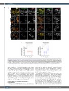
Figure 2. Cells expressing wild-type and mutated b1-tubulin demonstrate C-terminal polymodification. (A) Constructs carrying wild-type (WT), patient variants (p.R359W and p.L361Afs*19), and a C-terminal tail truncation of b1-tubulin fused to the fluorescent reporter mApple at the N-terminus were designed and cloned. (B) The WT construct was first transfected into Hek293T cells, which were then fixed and immunostained for polyglutamylated and polyglycylated tubulin residues specifically. (C and D) A comparison of the WT and mutated constructs shows a reduction in polyglutamylation and polyglycylation in each mutant compared to the WT. (n=3 independent differentiations, standard deviation plotted on graphs. Two-way ANOVA with multiple comparisons performed to establish significance.)
when compared to WT platelet forming iPSC-MK (Figure 3K). While polyglycylated and polyglutamylated residues form a distinct peripheral band around WT MK as shown in Figure 3K, the KO cells demonstrate a diffuse tubulin staining (evidenced by line profiles in Online Supplementary Figure S7). Figure 3L shows quantification of this staining whereby WT iPSC-MK display higher mean intensity than KO constructs in polyglycylated tubulin, however no significant quantitative change in polyglutamylation was observed. This is the first data demonstrating the effects of the loss of TUBB1 expression in human MK.
Platelets demonstrate a different pattern of polymodification
We hypothesized that the b1-tubulin polymodifications
evident in MK might be differently regulated between resting and activated platelets. We therefore compared immunofluorescence staining of polyglutamylated and polyglycylated tubulin between resting platelets and cells spread on fibrinogen and collagen.
Resting platelets demonstrate polyglutamylated tubulin which partially co-localizes with the b1-tubulin marginal band unlike MK which demonstrate extensive polyglycy- lation in proplatelet forming cells (Figure 4A). On fibrino- gen and collagen spreading, polyglutamylation is evident, notably at the marginal band of spreading cells on fibrino- gen (Figure 4B). A lack of polyglycylation is consistent in both resting and activated platelets. Western blotting of resting platelets and cells activated through stimulation by CRP over time does not show an increase in the total
248
haematologica | 2022; 107(1)
