
Page 237 - 2022_01-Haematologica-web
P. 237
Baseline SUVmax in FL and tumor proliferation
AB
C
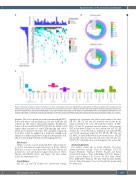
Figure 6. Molecular profiling of follicular lymphoma samples. (A) Heatmap of the most significantly mutated genes in follicular lymphoma (FL) samples from the training cohort (one column by sample, one line by gene). The colors refer to the type of mutation. (B) Circos plot illustrating the functional pathways involved in FL genomic abnormalities in the training (upper panel, n=33) and validation (lower panel, n=18) cohorts. The percentages refer to the frequency of alterations in the respective pathways. (C) Histogram showing the frequency of impaired functional pathways according to whole body maximum standardized uptake (SUVmax) levels.
patients. We also found an association between high SUVmax levels and tumor cell proliferation, but not with the cell content in the tumor microenvironment. Proliferative tumor cells are enriched in FOXO1 mutations, which could explain their resistance to anti-CD20 therapy and subse- quent poorer patient outcomes. The integrative approach used here could be applied in a multi-site lymph node analysis to further clarify the biological heterogeneity of FL and its relationship with functional imaging patterns.
Disclosures
CR has received a research grant from Roche and personal fees as well as non-financial support from Janssen, Roche, Takeda; ROC has received research grant from Gilead and Takeda and personal fees and non-financial support from Janssen, Roche, Takeda, Merck/BMS, Abbvie and Amgen. The other authors declare no competing interests.
Contributions
CR, PG, CL and CB designed the experimental strategy,
organized the experiments and collected and analyzed the data; CR, PG, PB, LY, LO and CG provided clinical data for the patients included in the series; SK performed analyses of PET- FDG; TF and JG performed statistical analyses; MT generated SES and performed analysis by data mining with CR, CB, JJF and CL; CL selected FL biopsies, performed and analyzed IHC and t-NGS experiments helped by PG, SP, SE, SR, LM, and CS; ROC, NA and DMF provided experimental advice; CR, JJF, CL and CB wrote the manuscript with advice from AS, CK, CL, MT, LY, PG. All authors discussed and approved the manuscript.
Acknowledgements
The authors would like to thank Nathalie Van-Acker (immunochemistry staining, Dept. of Pathology, IUCT-O, Imag’in Plateform, Toulouse), Frédéric Escudié and David Grand (NGS analysis, Dept. of Pathology, IUCT-O, Toulouse, Karin Gordien (Registre des Cancers du Tarn, Albi), Sophie Péries (CRB CHU Toulouse). We also thank Suzanne Rankin from the Dijon-Bourgogne University Hospital for proofreading the manuscript.
haematologica | 2022; 107(1)
229
