
Page 211 - 2022_01-Haematologica-web
P. 211
Phenogenomic features of post-transplant PBL
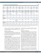
Table 1. Clinical features of post-transplant plasmablastic lymphomas.
Patient 1 2 3 4 5 6 7 8 9 10 11
Age,sex 61,M 70,M 76,M 65,M 22,M 12,F 72,M 57,F 64,M 15,M 60,F
Indication for tx
ETOH cirrhosis
NIDCM Heart
Cyclosporine, Prednisone
Small intestine, pulmonary valve
CAD Heart
Cyclosporine, Prednisone
Nasal
PCKD Kidney
Cyclosporine, Prednisone
Skin, spinal epidural tissue
Obstructive uropathy
Kawasaki disease
IPF LAM Lung Lung
Alport syndrome
HLHS
Pulmonary fibrosis
Type of tx
Immuno- Azathioprine. MMF, Azathioprine, MMF, Cyclosporine, Azathioprine, Azathioprine, Azathioprine, Azathioprine, Cyclosporine, MMF,
Liver and kidney
Kidney Prednisone
Heart
Tacrolimus, Prednisone
Small intestine, pleura
Kidney
Tacrolimus, Prednisone
Small intestine, pelvic soft tissue
Heart Prednisone
Lung
Tacrolimus, Prednisone
Liver
suppression at diagnosis
Site of involvement
Marrow involvement
Stage
Time to development
of PTLD (years)
Therapy
Dx to last
follow up ( years) and status
Cause of death
Tacrolimus, Prednisone
Pleural and peritoneal fluid
Skin, LN
Tacrolimus, Prednisone
Small intestine, peritoneal fluid
Tacrolimus, Prednisone
Small intestine
Small intestine
NA
IV IV I IV IV IV IV IV IV IV I
0.6
None 0.03, dead
PTLD/sepsis
Neg 7.0
Neg 11.1
Neg 1.3
Neg 7.8
Neg 10.3
NA 11.9
NA 0.9
R-EPOCH 0.4, dead
ICH
Neg 9.6
Neg 10.8
NA 10.7
R-EPOCH, Rituximab
1.4, dead
Sepsis
R-EPOCH 0.5, dead
Unknown
Radiotherapy 4.1, dead
Cardiac arrest
Radiotherapy, Bortezomib- Dexamethasone
5.4, dead
PTLD
R-CP, FCM 0.3, dead
PTLD
CP, GemOx, Surgery
15.9, alive
NA
R-EPOCH 2.0, dead
PTLD
None 0, dead
Sepsis
Bortezomib- EPOCH, ASCT
6.0, alive
NA
M: male; F: female; tx: transplant; dx: diagnosis; ETOH: alcohol-related; NIDCM non-ischemic dilated cardiomyopathy; CAD coronary heart disease; PCKD: polycystic kidney disease; IPF: idiopathic pul- monary fibrosis; LAM: lymphangioleiomyomatosis; HLHS: hypoplastic left heart syndrome; MMF mycophenolate mofetil; LN lymph node;; PTLD: post-transplant lymphoproliferative disorder; R-EPOCH: rituximab, etoposide, vincristine, doxorubicin, cyclophosphamide, prednisone; R-CP: rituximab cyclophosphamide, prednisone; FCM: fludarabine, cyclophosphamide, mytoxantrone; CP: cyclophos- phamide, prednisone; GemOx: gemcitabine, oxaliplatin; ASCT autologous stem cell transplant; Dx: diagnosis; NA: not analyzed; ICH: intracranial hemorrhage.
seropositive for EBV at the time of transplantation. EBV viremia at diagnosis was observed in all four EBV+ and two of five patients with EBV– PBL with available results (Online Supplementary Table S2).
IGH gene rearrangement analysis
All PT-PBL and both preceding monomorphic PTLD
showed clonal IGH gene rearrangements. Clonal relatedness was established in all three recurrent PBL and between the PBL and prior monomorphic PTLD. The polymorphic PTLD demonstrated oligoclonal products.
Cytogenetic abnormalities
Cytogenetic findings are listed in Table 3. All three PBL with informative results showed complex karyotypes. IGH- MYC rearrangements were detected in five of 11 (45%) cases (3 at diagnosis, 2 at recurrence). Multiple copies of MYC due to polyploidy were detected in two cases (concur- rent with MYC rearrangement in 1 case). Three of six cases with MYC abnormalities (2 with rearrangements, 1 with gain) showed ≥40% MYC positivity by immunohistochem- istry. PBL of patients who died of PTLD, and those who did not, revealed MYC abnormalities in two of four (50%) versus four of seven (57%) cases, respectively (P=1.0). Two cases demonstrated IGH rearrangements with unknown partners. Chromosome 17/TP53 abnormalities were detected in six; subclonal (30-40% of cells) monosomy 17 (1 EBV+, 1 EBV–), multiple copies (polyploidy) of chromosome 17 (2 EBV–), and TP53 deletion (1 EBV+, 1 EBV–). Loss of TP53 was detect- ed by targeted genomic sequencing in two cases, including one case with insufficient material for FISH analysis.
No MYC or TP53 alterations were observed in the poly-
morphic PTLD or monomorphic PTLD (DLBCL) preceding PBL. Insufficient material precluded analysis of the PT-plas- macytoma.
Targeted genomic sequencing
Pathogenic and likely pathogenic somatic non-synony- mous single nucleotide variants (SNV) are listed in Table 3 and all, including variants of unknown significance (VUS), are listed in Online Supplementary Table S1. Excluding sam- ples from two patients with high-level MSI, which exhibited up to 194 total SNV, the PBL harbored one to 16 pathogen- ic/likely pathogenic (median 7) and three to 26 total SNV (median 13) including VUS. MSI status was confirmed by a polymerase chain reaction-based method. All four PBL sam- ples classified as having high-level MSI (from patients 6 and 7) demonstrated loss of expression of at least two mismatch repair (MMR) proteins (Table 2). After factoring out cases with high level MSI, the number of variants was still higher in EBV– than EBV+ PBL, but the difference was not statistical- ly significant (4.7 mean pathogenic/likely pathogenic and 9.3 total SNV in EBV+ vs. 10.0 mean pathogenic/likely pathogen- ic and 19.3 total SNV in EBV– cases; P=0.15 and P=0.09, respectively). There was no a statistically significant differ- ence in the number of variants between patients who died of PBL and those who did not (4.7 mean pathogenic/likely pathogenic and 10.0 total SNV vs. 7.3 mean pathogenic/like- ly pathogenic and 14.0 total SNV; P=0.51 and P=0.6, respec- tively).
In our series, mutations were most frequent in epigenetic modifier genes, occurring in eight of 11 (73%) patients, and included recurrent mutations in the KMT2/MLL family of methyltransferases (KMT2C, n=5; KMT2D, n=3; and
haematologica | 2022; 107(1)
203
