
Page 206 - 2022_01-Haematologica-web
P. 206
T. Braun et al.
Our computational analyses of mRNA targeted by dereg- ulated miR in T-PLL suggest a strong impact of altered miR clusters on activation, death resistance, and aberrant DNA damage responses. Abnormal activity of these pathways, triggered by TCL1A overexpression and damaging ATM aberrations, has emerged as a hallmark of T-PLL
Table 2. Prognostic score (miROS-T-PLL) including miR-200a-3p, miR- 223-3p, and miR-424-5p expression levels.
pathobiology.1,11 We propose that protumorigenic miR net- works in their function as posttranscriptional regulators may further enhance the effects of these key genomic lesions, contributing substantially to the pathogenesis of T- PLL. Similar cooperative miR-mRNA networks were postu- lated for T-ALL and CLL.35,45 The causes of the miR deregu- lations we observed here, remain unknown and are, besides TCR activation, likely multifactorial. As T-PLL is character- ized by a strong genomic instability and high burdens of reactive oxygen species,1,46 mutations or copy number alter- ations of miR-encoding genes provide possible explana- tions, in addition to epigenetic mechanisms. Notably, inci- dences of genomic losses of the downregulated miR-140- 3p, miR-196b-5p, miR-339-3p, and miR-589-5p were in the order of 4-9% of T-PLL cases in our cohort.
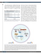
In summary, we identified a T-PLL-specific miR-ome, with 34 differentially deregulated miR, that appears instructed by TCR activation. By integrating altered miR expression with the information derived from transcrip- tome analyses, we postulate that the miR-ome of T-PLL shapes (dys)regulated networks towards apoptotic resist- ance, cell cycle abrogation, and defective DNA damage repair (Figure 6). Highlighting the pathobiological impact of the discovered miR deregulations, we developed the first
Parameter
miR-200a-3p expression1
(FC, rel. to healthy CD3+ pan-T cells)
miR-223-3p expression1
(FC, rel. to healthy CD3+ pan-T cells)
miR-424-5p expression1
(FC, rel. to healthy CD3+ pan-T cells)
Prognostic Groups
Lower Risk Higher Risk
0 Points
≥ 2.21 < 9.80 < 0.91
0-1 points 2-3 points
1 Point
< 2.21 ≥ 9.80 ≥ 0.91
1evaluated by small-RNA sequencing and compared to the mean expression of CD3+ pan-T cells of six healthy donors: FC: fold change; miR: microRNA; T-PLL: T-cell prolym- phocytic leukemia; miROS-T-PLL: miR-based overall survival score for T-PLL.
198
haematologica | 2022; 107(1)
Figure 6. Graphical summary of postulated microRNA/mRNA-based deregulated networks in T-cell prolymphocytic leukemia. T-cell activity shaped microRNA expression signatures (miR-omes)/ transcriptome networks are displayed as identified by our combinatorial approach of small-RNA and transcriptome sequencing analyses in 41 clinically well characterized T-cell prolymphocytic leukemia (T-PLL) cases. MiR differentially expressed in T-PLL and mRNA associated with these miR (P<0.05) are presented. The background color of miR and mRNA (dots) indicates the fold changes of differential expression as compared to age-matched healthy donor-derived CD3+ pan-T cells (blue=downregulation, red=upregulation). MiR-223-3p, the miR-21 family, the miR-29 family, and the miR-200c/141 family emerged as hallmarks of the T-PLL miR-ome, as they were (i) significantly deregulated among T-PLL cases, (ii) presented putative targets involved in oncogenic pathways of T- PLL’s pathobiology, (iii) showed associations with prognostic parameters, and (iv) were already described in the leukemogenesis of other B- and T-cell malignancies. Deregulations of these four miR-families were associated with cooperative effects on DNA damage response pathways as well as on pro-proliferative and cell survival signaling (as revealed by gene set enrichment analysis based on correlated mRNA). Genes previously described as hallmarks of T-PLL (e.g., TCL1A, CTLA4, and MYC) were found within the network of the identified miR-associated mRNA.
