
Page 18 - 2022_01-Haematologica-web
P. 18
G. Cartron and J. Trotman
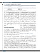
Table 3. Adverse prognostic factors according to FLIPI, FLIPI-2 and PRIMA-PI.
FLIPI30
Age > 60 years Ann Arbor stage III/IV LDH > normal Hemoglobin < 120 g/L
Nodal sites involved > 4
FLIPI-231
Age > 60 years Marrow involvement b2-microglobulin > normal Hemoglobin < 120 g/L
Tumor mass > 6 cm
PRIMA-PI20
Marrow involvement b2-microglobulin > 3 mg/L
FLIPI: Follicular Lymphoma International: Prognostic Index; PRIMA-PI; PRIMA Prognostic Index; LDH: lactate dehydrogenase.
been validated in studies using immunochemotherapy and is now widely used in daily practice and clinical trials. FLIPI-2 was developed in a population of rituximab-treat- ed patients for whom b2-microglobulin was available31 and is also predictive of PFS and OS. The PRIMA prog- nostic index (PRIMA-PI) was developed from a retrospec- tive analysis of a population of patients receiving immunochemotherapy followed by rituximab mainte- nance25 in a large randomized clinical trial. This prognos- tic index is based on b2-microglobulin and marrow infil- tration, two easily available parameters, albeit with the requirement of an invasive bone marrow biopsy. Recently, with the description of recurrent gene muta- tions in FL, some groups have designed new prognostic indices, including both clinical factors and mutational sta- tus.32,33 The use of these indices is uncommon in routine practice due to the limited availability of mutational analysis, and they have only been validated as prognostic in the FL population treated with R-CHOP (rituximab plus cyclophosphamide, doxorubicin, vincristine, pred- nisone).
Natural course of the disease
FL is still considered as an indolent but usually incur- able disease, although the age- and gender-matched sur- vival in patients who have remained event-free for 2 years after initial treatment challenges our concepts of cure.34 FL is characterized by a heterogeneous presenta- tion and outcome reflecting its biological heterogeneity. Patients with advanced stage indolent disease are man- ageable with a “wait and watch” strategy for many years whereas others may experience short OS related to trans- formation into diffuse large B-cell lymphoma. In the for- mer population, representing one third of patients, an indolent presentation does not require therapeutic inter- vention.35 For such asymptomatic patients, 50% and 20% will not require treatment at 3 and 10 years, respectively, after diagnosis and their estimated OS is probably higher than the median 80% charted at 10 years after rituximab chemotherapy.1,2,28 The pattern of clinical evolution seems to be the same for FL grades 1,2 and 3A whereas, grade 3B FL is now recognized to be genetically closer to diffuse large B-cell lymphoma with a more aggressive course that requires anthracycline-containing immunochemotherapy at diagnosis.36 For patients requiring treatment, the use of combined immunochemotherapy results in a 10-year OS of around 80%.2 However, the lack of a plateau in the PFS curve suggests that we have probably only extended the natural history of this indolent disease with repeated relapses at increasingly shorter intervals. Despite recent therapeutic progress and improved OS, FL remains the leading cause of death in patients with a cumulative inci- dence of 10% at 10 years.3 Nonetheless, even after first
relapse/progression the survival data are promising, with a median OS for patients who received second-line treat- ment beyond 10 years.37 Patients experiencing disease progression within 24 months after treatment initiation (POD24) represent a particular group of need, with a 5- year survival of only 50%.38 However, it should be noted that this datum was derived in an era that predated PET- based staging and in one large retrospective population- based study the prognostic impact of POD24 is not as powerful in the modern era.39
Histological transformation is probably a turning point for patients’ outcome with a shorter survival if it occurs after FL treatment. De novo histological transformation (i.e., diffuse large B-cell lymphoma histology with patho- logical findings showing existence of a FL component at the time of diagnosis) has a better prognosis.40 The medi- an survival after histological transformation is around 4 to 5 years and probably shorter if the transformation occurs within the first year.41 The annual incidence of biopsy- documented histological transformation is <3%, with a plateau after the first 2 years following treatment initia- tion.11,41 One study of more than 8,000 patients identified a lower incidence of histological transformation in the rit- uximab era. The 10-year cumulative hazard of histologi- cal transformation was 5.2% (95% CI: 4.5-6.2) in patients who received rituximab and 8.7% in those who did not (hazard ratio=0.73, 95% CI: 0.58-0.90; P=0.004).42 Histological transformation is probably under-reported given that biopsy, while recommended, is not uniformly performed at relapse/progression.
Initial treatment of follicular lymphoma
After distinguishing localized from disseminated disease applying the Lugano 2014 classification, the GELF/BNLI28,29 (Tables 1 and 2) treatment-initiation indices help to identi- fy patients with symptomatic FL requiring treatment. A combination of staging and GELF/BNLI characteristics dis- tinguishes three clinical situations that require different therapeutic approaches: (i) patients with localized FL; (ii) patients with disseminated FL not meeting treatment-initi- ation criteria (also called “low-tumor burden”); and (iii) patients with disseminated FL who meet treatment-initia- tion criteria (also called “high-tumor burden”).
Localized follicular lymphoma
Fewer than 20% of patients present with stage I or II FL at diagnosis.37 Such patients are identified after a compre- hensive assessment, including both PET and bone marrow biopsy to confirm the localized nature of the disease. Stage I disease, with removal of the only pathological node (stage I0) is an infrequent clinical situation that can benefit from therapeutic abstention. While data are sparse, addi- tional treatment (such as radiotherapy) does not appear to
10
haematologica | 2022; 107(1)
