
Page 17 - 2022_01-Haematologica-web
P. 17
Individualized first-line management of FL
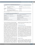
Table 1. Revised staging system for primary lymphoma (adapted from Cheson et al.76).
Stage
Limited I
II
I or II bulky*
Advanced III
IV
Nodal involvement
One or several nodes of one group
Two or more nodal groups on the same side of the diaphragm
I or II as above with bulky disease
Nodes on both sides of the diaphragm or nodes
above the diaphragm with spleen involvement
Additional non-contiguous or extended extranodal involvement
Extranodal involvement (E)
Single extranodal
Stage I or II with contiguous and limited extranodal involvement
Not applicable
Not applicable Not applicable
*‘Bulky’disease has been defined as any nodal mass of 10cm or greater than one third of the transthoracic diameter at any level of thoracic vertebrae.Tonsils,Waldeyer ring and spleen are considered nodal sites.
Table 2. Criteria for initiation of treatment in patients with follicular lymphoma.
GELF criteria27
Presence of at least one of the following criteria:
Any B symptom
Involvement of ≥3 nodal sites, each with a diameter ≥ 3 cm Tumor mass ≥7 cm
Symptomatic splenomegaly
Pleural effusion or ascites
Organ compression
Serum LDH or b2M above upper limit of normal
GELF: Groupe d'Etude des Lymphomes Folliculaires; BNLI: British National Lymphoma Investigation; LDH: lactate dehydrogenase; b2M: beta-2 microglobulin.
BNLI criteria28
Presence of at least one of the following criteria:
Pruritus or B symptom(s)
Rapid generalized disease progression in the preceding 3 months Life-endangering organ involvement
Significant bone marrow infiltration
Localized bone lesions detected on X-ray or isotope scan
Renal infiltration
‘Macroscopic’ as opposed to ‘microscopic’ liver involvement
tests, lactate dehydrogenase and b2-microglobulin), CT- scan of the neck, thorax, abdomen and pelvis and PET.
PET is now the recommended gold-standard imaging investigation at initial staging. FL has a universally, albeit not uniformly, glucose-avid histology, with FDG uptake in 98% of patients.15 PET is more sensitive than standard contrast-enhanced CT at detecting extranodal disease.16 The PET report should indicate nodal and extranodal 18F- FDG uptake due to lymphoma as well as the lesion with the highest standardized glucose uptake value (SUVmax). The extent of the disease is then classified according to the Lugano classification (Table 1). PET scanning resulted in up-staging of as many as 62% of localized (I/II) cases into advanced stage (III/IV) cases.17 The utility of PET has been demonstrated by identifying disseminated disease ultimately associated with a poorer outcome if treated as localized on the basis of CT-based staging.17 The prognos- tic value of total metabolic tumor volume at diagnosis in FL remains debated12,18 and the predictive value of auto- mated software solutions for measuring total metabolic tumor volume19 needs to be validated in prospective stud- ies before being utilized in a standard manner in clinical practice. The prognostic value of end-of-induction PET response after first-line immunochemotherapy for FL20,21,22 is now clear and provides a platform for PET-adapted therapies in current clinical trials.23,24
While focal bone marrow involvement can be identi- fied on PET, it is generally less sensitive than bone mar- row biopsy because it is often diffuse and low volume. Thus, when identification of bone marrow involvement is necessary (cytopenia, clinical trial, confirmation of localized disease), bone marrow biopsy (including both aspirate and trephine) is required to complete marrow assessment when the PET is negative. Identification of marrow involvement does not change treatment in dis-
seminated disease and its prognostic value is debated, with prognostic merit identified in the PRIMA study25 but not in the GALLIUM study.26
Treatment-initiation criteria
Tumor burden is usually considered an important prog- nostic factor in FL. This observation and the commonly indolent nature of FL led the Groupe d'Etude des Lymphomes Folliculaires (GELF)27 and the British National Lymphoma Investigation (BNLI)28 to propose cri- teria for initiation of treatment (Table 2). These are main- ly clinical criteria, empirically defined in the 1990s. They are still used to guide initial management in both clinical practice and trials and have shown consistent prognostic value.29 Notwithstanding the value of such criteria, in the patient with a slowly waxing and waning disease, it may not be appropriate to commence treatment in routine clinical practice just because they have a mildly elevated lactate dehydrogenase or an asymptomatic abdominal mass that has taken years to reach 7 cm in longest dimen- sion. Conversely, some young patients who do not strict- ly meet GELF/BNLI criteria may warrant initiation of therapy for rapidly progressing disease.
Prognostic indices
In 2004, the Follicular Lymphoma International Prognostic Index (FLIPI) was the first prognostic index dedicated to FL30 (Table 3). FLIPI separates three groups of patients (approximately one third each) with significant differences in overall survival (OS) (Table 4). Although originally designed in the pre-rituximab era, FLIPI has
haematologica | 2022; 107(1)
9
