
Page 168 - 2022_01-Haematologica-web
P. 168
T. Wang et al.
nisms of Nupr1 in regulating HSC, we performed RNA- sequencing analysis of Nupr1-/- HSC from 8-week-old Nupr1-/- mice. Analysis of gene expression indicated that there were 319 genes differentially expressed between WT
and Nupr1-/- HSC (>2-fold difference in expression; adjusted P value <0.05 [DESeq2 R package]). Gene-ontology analysis of these differentially expressed genes indicated enrichment of genes involved in regulation of mitotic cell cycle and neg-
AB
C
D
E
F
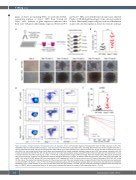
Figure 4. Deletion of Nupr1 promotes hematopoietic stem cell expansion in vitro. (A) Schematic diagram of hematopoietic stem cell (HSC) expansion in vitro. Fifty CD150+KSL HSC from wild-type (WT) and Nupr1-/- mice were sorted into fibronectin-coated plate wells, containing albumin-free F12 medium supplemented with 1 mg/mL polyvinyl alcohol (PVA), 100 ng/mL thrombopoietin (TPO) and 10 ng/mL stem cell factor (SCF). HSC were cultured for 10 days and then analyzed by flow cytom- etry (FACS). For the limiting dilution assay, serial doses were transplanted into lethally irradiated recipients, together with 2×10⁵ bone-marrow competitor cells. (B) Number of cells derived from 50 HSC after 10 days of culture in vitro. Data were analyzed using an unpaired Student t-test (two-tailed) and are represented as mean ± standard deviation (SD) (WT, n=10; Nupr1-/-, n=16). ***P<0.001. (C) Representative images of WT and Nupr1-/- HSC from freshly isolated HSC (day 0) and after 10 days of culture (day 10). Images of five representative colonies (biological replicates) are shown. (D) Representative flow cytometric plots of HSC from cultured WT and Nupr1-/- HSC at day 10. p-HSC: primary HSC from bone marrow. e-HSC: expanded HSC after 10 days of culture ex vivo. (E) Counts of phenotypic CD150+KSL HSC at day 10 after culture. The dashed line indicates the amount of the primary input cells. Data were analyzed using an unpaired Student t-test (two-tailed) and are represented as mean ± SD (WT, n=8; Nupr1-/-, n=11). **P<0.01. (F) Poisson statistical analysis after limiting-dilution analysis; plots were obtained to allow estimation of competitive repopulating units in each condition (n=10 mice transplanted at each dose per condition, *P<0.05). The plot shows the percentage of recipient mice containing less than 1% CD45.2+ cells in the peripheral blood at 16 weeks after transplantation versus the number of cells injected per mouse. *P<0.05.
160
haematologica | 2022; 107(1)
