
Page 109 - 2022_01-Haematologica-web
P. 109
RNA-sequencing of fusion genes in AML
aberrant transcription or trans-splicing events. Many fusion genes have been described as drivers across multiple human cancer entities.1 Hematopoietic malignancies are well-char- acterized regarding the abundance of fusion genes, e.g, chronic myeloid leukemia harbors the BCR-ABL1 fusion in more than 95% of cases, and acute promyelocytic leukemia is characterized by PML-RARA in 90% of cases. In acute myeloid leukemia (AML), fusion genes are found in about 30% of patients2 and are often regarded as major markers, defining clinically relevant subtypes.3-6 Their identification is crucial for risk assessment and deciding treatment strate- gy. During the initial diagnosis of AML, fusion genes are detected using conventional metaphase karyotyping (here- after referred to as Karyotyping) and/or targeted molecular assays (hereafter referred to as molecular diagnostics) such as fluorescence in situ hybridization (FISH) or reverse-tran- scriptase polymerase chain reaction (hereafter referred to as PCR). On a chromosomal level, Karyotyping detects abnor- malities by light microscopy of metaphase spreads, where- as FISH labels chromosomal alterations using specifically designed probes that bind to particular genomic regions of interest. On a molecular level, PCR may confirm the pres- ence of a specific genomic or transcriptomic sequence by targeted amplification. However, these methods are labori- ous and time-consuming, depend on the experience of the analyst and might be subject to erroneous assessments. Furthermore, the resolution of Karyotyping is limited to the microscopic level of chromosomal arms/bands and PCR/FISH can only be used to analyze predefined targets. Small inversions, duplications or short interstitial deletions as well as cryptic fusions are difficult to detect with these procedures. Although FISH and PCR are suitable for the tar- geted detection of submicroscopic lesions, they are not rou- tinely applied to the systematic identification of previously uncharacterized aberrations and usually serve as comple- mentary validation methods.
Over the last decade, next-generation sequencing tech- niques have evolved tremendously and are being increas- ingly used in clinical diagnostics.7-9 Next-generation sequencing methods enable scalable genomic analyses, ranging from single genes and gene sets of interest up to
genome-wide analyses, covering the entire genome at sin- gle base pair resolution. Furthermore, RNA-sequencing enables transcriptome-wide studies, covering all transcribed genes present in a cell. Recently, a study proposed a single bioinformatic pipeline for AML diagnostics which inte- grates detection of fusion genes, small variants, tandem duplications and gene expression from RNA-sequencing data.10 Thus, DNA and RNA-sequencing allow for the examination of a wide range of genetic lesions, including the discovery of novel aberrations. Sequencing technologies are improving quickly and innovation in this field continu- ously reduces time and costs for genomic analyses, which enables the processing of even more samples in parallel with even greater precision. Simultaneously, developments in computational biology can exploit these advancements for accurate detection of genetic aberrations.
To date, more than 20 algorithms for fusion gene detec- tion by RNA-sequencing have been published9,11,12 but iden- tification of fusions using RNA-sequencing remains chal- lenging and a high rate of false positives is common. Therefore, careful evaluation of fusion calls and appropriate filtering strategies are needed to enable reliable application of this technology in diagnostics. In AML, no comprehen- sive comparison of fusion gene detection by RNA-sequenc- ing and clinical routine has been reported so far. In this study, we set out to assess the potential of RNA-sequencing for the detection of clinically relevant fusion genes in com- parison to standard diagnostic methods. Additionally, we developed several filters for robust fusion gene identification and the discovery of novel rearrangements in AML patients.
Methods
Patients’ samples
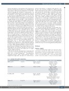
A total of 806 AML patients’ samples were subjected to whole- transcriptome sequencing. The samples were collected from with- in four different cohorts: (i) the German AML cooperative group (AMLCG 2008 and AMLCG 1999, n=257);13,14 (ii) the German Cancer Consortium (DKTK, n=69);15,16 (iii) the Beat AML program (n=423);17 and (iv) the Institute for Molecular Medicine Finland
Table 1. Summary of the patients’ characteristics.
Cohort
AMLCG (n=257)
DKTK (n=69)
Beat AML (n=423)
FIMM (n=57)
Median age (range)
58 (18-79)
61 (21-85)
61 (2-87)
58.5 (21-77)
Sex, n (%)
Females = 131 (51.0) Males = 126 (49.0)
Females = 31 (44.9)
Females = 186 (44.0) Males = 237 (56.0)
Females = 29 (50.0) Males = 29 (50.0)
ELN risk group, n (%)
Favorable = 75 (29.2) Intermediate = 61 (23.7) Adverse = 107 (41.6) NA = 14 (5.5)
Favorable = 33 (47.8) Males = 38 (55.1) ntermediate-I = 25 (36.2) Intermediate-II = 7 (10.1) Adverse = 3 (4.4) NA=1(1.5)
Favorable = 112 (26.5) Intermediate = 141 (33.3) Adverse = 148 (35.0) Favorable or Intermediate = 13 (3.1) Intermediate or Adverse = 7 (1.6) NA=2(0.5)
Favorable = 9 (15.8) Intermediate = 19 (33.3) Adverse = 18 (31.6) NA = 11 (19.3)
ELN: European LeukemiaNet; NA: not available.
haematologica | 2022; 107(1)
101
