
Page 197 - 2021_12-Haematologica-web
P. 197
Letters to the Editor
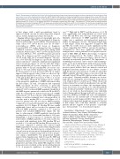
Figure 1. Reassessment of traditional risk factors in first multiple myeloma relapse shows improved prognostic accuracy compared to their evaluation at diag- nosis (shown in the Online Supplementary Appendix). GEP70 high-risk (HR) patients at relapse had significant worse progression-free survival (PFS) (A), and OS (B) compared to low-risk (LR) patients, P<0.01. HR fluorescence in situ hybridization (FISH) included translocation t14;16, t4;14 and del 17p and showed worse PFS (P=0.1) (C) and OS (P<0.05) (D), compared to patients with LR FISH. Assessment of revised International Staging System (RISS) at relapse showed significant worse PFS, E, and OS, F, for patients with RISS 2+3 compared to patients with RISS stage I. The presence of >3 focal lesions by positron emission tomography and computed tomography (PET CT) at relapse was associated with worse PFS (H) and OS (I) in first relapse. The results were not significant, likely due to the relative small patient number. Achievement of minimal residual disease (MRD) negativity after first relapse was a powerful marker for significantly improved PFS (J) and OS (K).
at first relapse with a mild non-significant trend to improved clinical outcome in earlier stages after relapsed disease (Online Supplementary Figure S2A and D).
Imaging with positron emission tomography and com- puted tomography (PET CT) was performed at diagnosis (n=120) and at relapse (n=111). Of the 120 patients in our study cohort, 69% (n=75) had at least one 18 F-fluo- rodeoxyglucose (FDG) avid lesion at diagnosis. Sequential PET CT studies during first line treatment confirmed resolution of PET avid lesions during initial treatment. At first relapse, 44.5% (n=50/112) had at least one lesion. Of these, 68% (n=34/50) had also presented with a FL at diagnosis and 46% (n=23/50) had at least one FL at the same site as at initial diagnosis. The pres- ence of >3 FL by PET at diagnosis, a previously identified adverse risk factor10 only had a small and non-significant adverse prognostic impact on outcome after first relapse with a median PFS of 1.4 years compared to 1.8 years for patients with 0-3 FL and a median OS of 3.9 years com- pared to 4.8 years (Online Supplementary Figure S3A and B). Reassessment of focal lesions by PET CT at relapse improved the prognostic value of this test, albeit not sig- nificantly (median PFS for 0-3 FL: 1.8 years vs. 1.0 year for >3 FL and median OS: 4.4 years for 0-3 FL vs. 2.1 years for >3 FL) (Figure 1H and I). In a further step we evaluat- ed the prognostic significance of MRD achievement after the first relapse. In total, 116 patients had sequential MRD assessment by flow cytometry, as previously described,9 after initiation of a second line therapy of which 47 (40.5%) achieved MRD negativity. Nearly all of the MRD-negative patients also achieved a CR (n=45/47), while the remaining two patients had achieved a VGPR. Achievement of a deep response with MRD negativity during second line treatment was a strong predictor of outcome with a median PFS to second relapse of 1.3 years for patients who did not achieve MRD negativity compared to not reached for patients who achieved MRD negativity (P<0.01) (Figure 1J). Median OS was equally significantly better and not reached for patients who achieved MRD negativity compared to 3.7 years for patients who remained MRD-positive (Figure 1K). The time to achievement of MRD negativity varied greatly, reaching from 0.6 to 3 years with a median of 1.02 years. Intriguingly, a slower response to treatment and later achievement of MRD negativity (>1.02 years, n=24), was associated with significant better PFS (median PFS not reached vs. 1.6 years) and OS (median OS not reached vs. 2.8 years) compared to patients who achieved rapid MRD negativity(<1.02 years, n=23) (Online Supplementary Figure S3C and D). In a final step, we evaluated the asso- ciation between aforementioned independent risk factors and the hazards of experiencing death as well as progres- sion using a multivariable Cox proportional hazards model (Online Supplementary Table S1A and B). FISH and RISS were excluded from the analysis due to the overall small patient number that was assessed at relapse. Age at progression and time from initial MM diagnosis to first progression were included, as they previously had shown to be of prognostic significance in relapsed MM dis-
ease.11,12 High risk by GEP70 and the presence of >3 FL were significantly and independently associated with worse PFS and OS as was older age at first progression. Similarly, achievement of MRD negativity after first relapse was a significant and independent prognostic marker for improved outcome. Though prolonged time to first relapse (TT1P) was suggestive of improved PFS and OS, the results were not quite significant in this cohort, suggesting that TT1P as a prognostic marker is determined by other more significant variables. Our study provides a strong rationale to incorporate reassess- ment of GEP, FISH and RISS at first relapse to improve clinical prognostication. We further underscore the importance of FL assessment at relapse, a practice that is currently inconsistently performed. The importance of identifying focal lesions can be vital to clinical manage- ment, as we show that patients with an increased num- ber of FL tend to have worse outcome and furthermore previous reports indicate that bone marrow content and peripheral myeloma markers do not always correlate with the presence of focal lesions.13 Lastly, we show that MRD negativity after first relapse is associated with sig- nificantly better PFS and OS, which is in line with a pre- vious report.8 Currently available therapies are increas- ingly effective, making the achievement of deep respons- es in relapsed disease a realistic goal.14 While the present study is limited by a relatively small patient size and dif- ferences in treatment at relapse, the investigated prog- nostic factors have been shown to be valid independent of the treatment modality.15 Furthermore, key character- istics of our study are that all patients were uniformly treated during upfront therapy and had been exposed to a protease inhibitor, an IMiD and stem cell transplanta- tion, which are all currently part of standard first line treatment in newly diagnosed MM. Taken together our study provides important insight into prognostic features at first relapse that could help clinicians to reclassify patients and also suggests to treat patients – if perform- ance status permits - to a deep clinical response.
David Baker,1* Milan Bimali,2,3* Luis Carrillo,4
Archana Sachedina,5 Daisy Alapat,4 Md Shadiqul Hoque,1 Mathew Kottarathara,1 Richa Parikh,1 Amani Erra,1
Angel A. Mitma,1 Pankaj Mathur,1 Yetunde Ogunsesan,1 Lakshmi Yarlagadda,1 Sravani Gundarlapalli,1
Sharmilan Thanendrarajan,1 Maurizio Zangari,1
Frits van Rhee,1 Guido Tricot1 and Carolina Schinke1
1Myeloma Center, Division of Hematology/Oncology, Winthrop P. Rockefeller Cancer Institute, University of Arkansas for Medical Sciences; 2Arkansas Children’s Nutrition Center, Arkansas Children's Hospital; 3Department of Biostatistics, University of Arkansas for Medical Sciences; 4Department of Pathology, University of Arkansas for Medical Sciences and 5Department of Radiology, University of Arkansas for Medical Sciences, Little Rock, AR, USA
*DB and MB contributed equally as co-first authors. Correspondence:
CAROLNA SCHINKE - cdschinke@uams.edu doi:10.3324/haematol.2021.278779
haematologica | 2021; 106(12)
3217
