
Page 195 - 2021_12-Haematologica-web
P. 195
Letters to the Editor
Predicting risk of progression in relapsed multiple myeloma using traditional risk models, focal lesion assessment with PET-CT and minimal residual disease status
Novel therapeutic strategies have dramatically increased the depth of response and survival rates in mul- tiple myeloma (MM), but the disease remains incurable in most patients because of eventual relapse.1 The timing and disease course of relapsed MM can be highly vari- able, and most often the presentation of the first relapse can give more information on disease biology and overall prognosis than parameters identified at diagnosis.2 The dynamic change of clinical parameters during the disease course has recently shown to significantly impact sur- vival in MM patients,3 underscoring that prognostic mod- els, such as fluorescence in situ hybridization (FISH), International Staging System (ISS), revised-ISS (RISS) and gene expression profiling (GEP) in addition to focal lesion (FL) assessment can be useful prognostic tools at initial diagnosis,4,5 even though they have not been fully vali- dated in the relapse setting. Furthermore, in contrast to newly diagnosed myeloma, it is unknown whether the depth of response after salvage therapy also affects long term outcome in relapsed disease.6 This is particularly true for the achievement of minimal residual disease (MRD) negativity, a powerful prognostic tool in newly diagnosed MM,7 even though its importance in relapsed disease has started to be elucidated only recently.8
In order to explore whether reassessment of initial prognostic markers at relapse increases accuracy in pre- dicting outcome after relapse and to determine whether MRD achievement after the first relapse improves out- come, we investigated 120 patients who relapsed after MM diagnosis and initial treatment on our total therapy (TT) 2-6 protocols between 2000-2016. All patients achieved a complete response (CR) and subsequently relapsed, with the first relapse occurring after January 2014, the time point at which MRD assessment by eight- color flow cytometry was established systematically at the University of Arkansas for Medical Sciences.9 The majority of patients received an immunomodulatory imide drug (IMiD)-based triplet as second line, either in combination with a proteasome inhibitor (64%) or a CD38 targeting monoclonal antibody (27%). The median time to first relapse after initial diagnosis and treatment was 5 years (range, 0.9-18 years) with a median follow up after first relapse of 1.57 years (range, 0.18-6.0 years). Patients’ characteristics are presented in Table 1.
GEP70 classified 17% as high-risk (HR) patients at diagnosis. The proportion increased significantly to 35% at relapse (P<0.05). While the diagnostic GEP70 classifi- cation retained significant prognostic value at relapse with HR patients having significantly worse PFS (median: 0.93 years vs. 1.86 years; P=0.03), and OS (median: 2.12 years vs. 5.01 years; P<0.01) (Online Supplementary Figure S1A and B), reassessment of GEP70 at relapse improved prognostic accuracy with a median PFS of 0.76 years for HR versus 2.15 years for low-risk (LR) patients, P<0.01, and a median OS of 1.87 years for HR, while LR patients had not reached their median OS, P<0.01, Figure 1A and B. Similarly, we saw that reassessment of FISH and RISS at relapse improved accuracy in outcome prediction over initial assessment at diagnosis. HR FISH alterations were characterized by translocations t(4;14) and t(14;16) and deletion 17p. In particular the proportion of patients with del17p increased significantly from 12.5% (n=11/88) at diagnosis to 28% (n=17/59) at relapse. Despite the rela-
tively small number of patients with FISH at relapse, reassessment of FISH improved the predictive accuracy for PFS (median: 1.16 years vs. 1.75 years; P=0.1) and OS (median: 2.86 years vs. 4.38 years; P<0.05) (Figure 1C and D) compared to assessment at diagnosis (Online Supplementary Figure S1C and D). Furthermore, reassess- ment of RISS at relapse, was a more accurate tool in pre- dicting PFS (median PFS RISS I: 1.8 years vs. median PFS RISS II/III: 1.15 years; P<0.05) and OS (median OS RISS I: not reached vs. median OS RISS II/III: 2.9 years, P<0.01) (Figure 1E and F), compared to RISS evaluation at diagnosis (Online Supplementary Figure S1E and F). Of note is that very few patients presented with RISS III at relapse, which is likely due to early detection of relapsing disease in most patients, and hence still a relatively small tumor burden with low b-2-microglobulin values and normal albumin and lactate dehydrogenase (LDH). In contrast to GEP70, FISH and RISS, we only saw a modest prognostic impact of ISS evaluation at diagnosis or even
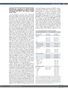
Table 1. Patient characteristics at diagnosis and relapse.
Age in yrs, (range) GEP high risk
ISS stage
1 2 3
FISH
Translocation t4;14 Translocation t14;16 Deletion 17p
R-ISS stage 1
2
3
Focal lesions by PET
0 1-3 >3
Treatment at relapse IMiD+PI
CD38 ab + IMiD CD38 ab + PI other#
Salvage ASCT at relapse+ Best response
sCR/CR VGPR PR
SD
PD
At diagnosis
59 (32-75) 20/115 (17.4%)
47/120 (39%) 39/120 (33%) 34/120 (28%)
11/84 (13%) 3/84 (3.75%) 10/84 (12%)
16/84 (19%) 54/84 (64.3%) 14/84 (16.7%)
39/114 (34%) 26/114 (23%) 49/114 (43%)
First line therapy 120/120 (100%)
At relapse
64 (37-81) 27/77 (35%)**
95/112 (84.8%)** 12/112 (10.7%)** 4/112 (3.8%)**
11/59 (18.6%) 3/59 (5%) 17/59 (28.8%)**
22/57 (38.6) * 32/57 (56%) 3/57 (5%)*
62/112 (55%)*
36/112 (32%)* 14/112 (12.5%)**
77/120 (64%) 32/120 (27%) 4/120 (4%) 6/120 (5%) 30/120 (25%) 2nd line therapy 60/119 (50.4%) 22/119 (18.5%) 17/119 (14.3%) 16/119 (13.4%) 4/119 (3.4%)
*P<0.05,**P<0.001 comparing presentation at relapse to diagnosis,McNemar’s test; #: regimen including intravenous chemotherapy such as cytoxan, adriamycin, etoposide, cisplatin (PACE, Metronomic, PACMED); +: salvage autologous stem cell transplant (ASCT) was performed in selected patients after brief reinduction with novel agents or intrvenous chemotherapy. GEP: gene expression profiling; ISS: International Staging System; FISH: fluorescence in situ hybridization; RISS: revised- ISS; PET: positron emission tomography; IMiD: immunomodulatory imide drug; PI: protease inhibitor; CR: complete remission;VGPR: very good partial remission, PR: partial response; SD: stable disease; PD: progressive disease.
haematologica | 2021; 106(12)
3215
