
Page 125 - 2021_12-Haematologica-web
P. 125
MCL-1 in human hematopoiesis
MCL-1 inhibitor S63845, together with increasing doses of the BCL-XL inhibitor A-1155463. Apoptosis induction was determined 24 h later (Figure 8A). Using SynergyFinder (https://synergyfinder.fimm.fi) a dose- response matrix was calculated (Figure 8B). The resulting Bliss score of 21.26 indicates strong synergy between the two inhibitors. Synthetic lethality was confirmed in colony-forming assays, both with cord blood- and bone marrow-derived CD34+ cells. Already at concentrations of 0.1 mM each, the drug combination resulted in a sub- stantial loss of colony-forming cells (compare Figure 8C, D with Figures 6B, C and 7).
To determine the number of immature cells with self- renewal potential that survived BCL-XL and/or MCL-1 inhibition, we used 10,000 cells isolated from primary colonies for serial colony-forming assays. Interestingly, only BCL-XL inhibition in the first plating resulted in depletion of progenitor cells able to form colonies in the
second plating. However, there was a synergistic effect when this BCL-XL inhibition was combined with MCL-1 inhibition (Online Supplementary Figure S5).
Discussion
Because of the narrow spectrum of cancer entities sus- ceptible to venetoclax, specific MCL-1 and BCL-XL inhibitors are eagerly awaited by oncologists. However, observations made in genetically modified mice indicate that inhibition of MCL-1 or BCL-XL could have more severe side effects than BCL-2 inhibition. Mice deficient for either MCL-1 or BCL-XL have severe developmental phenotypes while BCL-2-deficient mice lack lympho- cytes and melanocytes but are otherwise normal.10,40,41 Hematopoietic toxicity of anticancer drugs is responsible for most therapy-related morbidity and mortality and a
AB
CD
EF
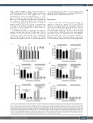
Figure 7. BCL-XL inhibition impedes colony formation and survival of erythroid cells. (A) Freshly isolated bone marrow was subjected to density gradient centrifu- gation and mononuclear cells were divided into CD34+ and CD34- cells using magnetic activated cell sorting technology. Both cell fractions were treated with the indicated concentrations of the BCL-XL inhibitor A-1155463. After 24 h, percentages of living cells were determined by flow cytometry using annexin V/7-AAD. Bars represent mean ± standard error of mean (SEM); n=3-4 from four independent experiments. (B-F) Human CD34+ cells, either derived from cord blood or bone mar- row, were differentiated in MethoCult culture containing 0.5 or 1.5 mM A-1155463. As controls, untreated and dimethylsulfoxide (DMSO)-treated cells were used (n=6-7 from seven independent experiments). After 11 days, total colony numbers (B) and total cell numbers (C) were determined using light microscopy and hemo- cytometry, respectively. Different cell types were determined by flow cytometry, and their absolute cell numbers were calculated (D-F). The following cell types were studied; immature CD34+ cells, monocytes (CD34-CD33+CD14+CD115-) and mature erythrocytes (CD71+CD235a+).
haematologica | 2021; 106(12)
3145
