
Page 62 - 2021_10-Haematologica-web
P. 62
B. Cieniewicz et al.
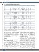
Table 2. Pediatric acute myeloid leukemia patient clinical characteristics
AML ID
3209 186 646 3281 3514 335 263 612 3491.1
3123 1355 794 683 882 351
1244
1563 3424 948 728
3082 758 258
Response to Sample LV-10 Timepoint
WHO Classification
AML - NOS
AML - NOS
AML with MDS-related AML - NOS
AML - NOS
AML - NOS
AML with mutated RUNX1 t(8;21); RUNX1-RUNX1T1 APL with PML-RARA
FAB Subtype
M5a
M5a M4Eo M5b
M2 M5a AML w/ MDS M2
M3
MPAL M3 M2 M4Eo MPAL M2
M7
M2 M5a M2 M1
M4Eo M4Eo MPAL
Cytogenetics % WBC Age Risk MRD
Sensitive
Intermediate Resistant
Onset Relapse Relapse Onset Onset Onset Onset Onset Onset
46, XY, t(X;11)(5'MLL+) 46, XY, MLL+
46, XY, del(7q)(q22)
46, XX
51, XY, +X, +9, +11, +14, +20 46, XY, FLT3-ITD+
46, XY
46, XY, t(8;21)
46, XX, t(15;17)
46, XX, Complex karyotype 46, XY, t(15;17)
46, XX, Inv(16)
46, XY, Inv(16)
46, XY, t(7;14)(q21;q32) 47, XX, t(8;21), +21
48, XY, +21, +Y
46, XX, t(8;21)
46, XX, t(9;11)
46, XX, Complex karyotype 46, XY, t(1;13)(p34~36;q13~14)[19] 46, XX, Inv(16)
46, XX, Inv(16)
46, XY
97 88 74 91 n/a 89 61 50 78
81 75 89 89 72 75
50
61 n/a 82 77
36 66 86
347.6 5 51.6 156 177 267
8.7 215 4 157 174 35 9.8 141 39.9 205 4.4 138
102 134 2.1 119 43 28 56.1 190
180.1 176 0.4 194
37 14
51.3 178 2.8 162 153.7 51 3.9 207
283 37 2.7 198 190 144
H - H + H + H - S + H - H + L - S -
H + S - L + L - H + S +
H +
L + S + H - H +
L - L - H -
Onset
Blast (103/mL) (M) group
Relapse MPAL
Resistant
Onset Onset Onset Onset Onset
Onset
Onset Relapse Onset Onset
Relapse Onset Onset
APL with PML-RARA Inv(16); CBFB-MYH11 Inv(16); CBFB-MYH11 MPAL
t(8;21); RUNX1-RUNX1T1, trisomy 21
AML with mutated RUNX1, trisomy 21 t(8;21); RUNX1-RUNX1T1 t(9;11); MLLT3-KMT2A AML with mutated NPM1 AML - NOS
Inv(16); CBFB-MYH11 Inv(16); CBFB-MYH11 MPAL
AML: acute myeloid leukemia; MRD: minimal residual disease after first induction chemotherapy; WBC: white blood cell; H: high; S: standard; L: low risk group. Pediatric acute myeloid leukemia (pAML) samples were grouped based on their sensitivity to LV-10-mediated killing.Sample timepoint,World Health Organisation (WHO) classification,French- American-English (FAB) classification, cytogenetics, blast percentage, white blood cell (WBC) count at diagnosis, age in months, risk group stratification, and minimal residual disease (MRD) status after first induction chemotherapy are displayed.
metric tests that do not assume equal variances between groups: Mann-Whitney or Wilcoxon test for groups of two (unpaired or paired samples, respectively), and Kruskal-Wallis or Friedman ANOVA with Dunn’s post hoc test for >2 groups (independent or dependent samples, respectively). Multiple testing correction was applied. Linear regressions were plotted using linear regression analysis in GraphPad Prism.
Results
Pediatric acute myeloid leukemia blasts have different levels of sensitivity to LV-10 killing
In order to determine if pAML can be killed by LV-10 cells, we first generated LV-10 cells from healthy donor- derived CD4+ T cells as described13,14 and verified their transduction efficiency, purity, cytokine profile, and killing capacity (Online Supplementary Figure S1). LV-10 cells had high transduction efficiency, high IL-10 and low IL-4, as well as high intracellular granzyme B expression at baseline (Online Supplementary Figure S1A to E) in comparison with effector T cell (Teff)-like control LV-GFP cells. LV-10 degran- ulation against target cells was also higher than LV-GFP cells, especially against HLA-class I positive myeloid tumor
cell lines U937 and ALL-CM (Online Supplementary Figure S1F). LV-10 cells were able to potently eliminate U937 and ALL-CM cells, but not HLA-class I negative ery- throleukemic K562 cell line (Online Supplementary Figure S1G). Target cell elimination was also observed in control LV-GFP cells, which are not tolerogenic13 and thus are not further explored for clinical use.
Next, we tested if LV-10 cells could eliminate pAML. We obtained 23 pAML bone marrow aspirates, 18 at onset and five at relapse, of various World Health Organization (WHO)29 and FAB diagnoses (Table 2). Killing-sensitive U937 and killing-resistant K562 cells were used as positive and negative controls, respectively. In the killing assay (see Materials and Methods), we observed three levels of pAML sensitivity to LV-10 killing: sensitive (S, >70% median elim- ination efficiency [E.E.]), intermediate resistant (IR, 25-70% median E.E.), and resistant (R, <25% median E.E.) (Figure 1A and B). Sensitivity or resistance was retested in nine pAML samples, and sensitivity levels were reproducible (not shown). Notably, all the pAML tested had high levels of HLA class I (not shown).
Because primary pAML typically expand poorly in vitro and can undergo spontaneous apoptosis, we examined if sensitivity correlated with pAML survival when cultured
2590
haematologica | 2021; 106(10)
