
Page 252 - 2021_10-Haematologica-web
P. 252
Letters to the Editor
AB
B
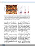
Figure 2. Time sequence of the appearance of red blood cell (RBC)-derived microparticles on the surface of human packed RBC isolated from (A) men and (B) women. Examples of atomic force microscopy (AFM) images with the use of WITec confocal CRM alpha 300 in non-contact mode (AC) (WITec, Ulm, Germany) and dry Zeiss objective (ECEPIPLAN 20x/0.4). AFM images of 256x256 lines and 512x512 lines were collected from areas of 25x25 mm2, 8x8 mm2 and 1.5x1.5 mm2, which were performed at room temperature on dried smears of (A) female RBC and (B) male RBC fixed with 1% glutaraldehyde (10 minutes). (C) Sex-spe- cific, time-dependent changes of the RBC-derived microparticle (RMP) sizes observed on the surface of human packed RBC. Data distribution is presented as box plots (median, Q1, Q3, interquartile range, min-max whiskers). Q1, Q3 indicate 25th and 75th percentiles, respectively. *Weeks 7 and 8 are additional meas- urements exceeding expiration date. Statistical significance of the obtained results (n>35) was tested with Kruskal-Wallis ANOVA non-parametric test followed by Tukey’s post hoc. NS: not significant; *P<0.05; **P<0.01, ***P<0.001, ****P<0.0001).
In the present study, we investigated the influence of donor’s sex on the sequence of changes observed during long-term storage of leukocyto-depleted pRBC contain- ing SAGM (saline, adenine, glucose, mannitol) additive solution and a trace amount of CPD (citrate, phosphate, dextrose) preservative, which were purchased from the Regional Center for Blood Donation and Hemotherapy in Krakow, Poland. According to the principles outlined in the World Medical Association Declaration of Helsinki, as well as a Bioethical Commission of the Jagiellonian University, venous blood for pRBC was obtained from volunteers including men (mRBC) n=12, aged <30 years (n=3), 30-39 years (n=4), >40 years (n=5) and women (fRBC) n=12, aged <30 years (n=4), 30-39 years (n=4), >40 years (n=4). All analyzes were carried out weekly throughout 6 weeks of pRBC storage, while the seventh and eighth week’s measurements were designed as addi- tional time points exceeding the pRBC expiration date (42 days), and were focused on the membranopathy on the level of pRBC membrane biochemistry, physical and mechanical properties (Figure 1), as well nanoscale changes (Figure 2).
Throughout the storage time, statistically significant differences between mRBC and fRBC in the kinetics of cholesterol and triglycerides’ increase were observed (Figure 1A and B). An increase between the first and eighth week was 2.27 times greater for cholesterol and 1.43 times greater for triglycerides in case of male donors. Such an increase in the lipid fraction in the sheathing solution during pRBC storage is related to lipidome alter- ations and disruption of phospholipid asymmetry in the RBC membrane.11 Our recent work and results of other groups suggest that the release of lipids from RBC mem- branes can be correlated with red blood cell-derived microparticle (RMP) formation.12 Our results agree with previous reports showing that the cholesterol level in fresh sample is similar in both sexes, while triglyceride level is lower in women.13,14
The levels of free iron, a renown and specific indicator
of hemolysis, were higher in mRBC, suggesting greater hemolysis rate (Figure 1D), what agrees with previous results.4,10 Lactate dehydrogenase (LDH) levels in both males and females below 40 years old did not differ, but higher average levels (Figure 1C) of LDH in fRBC could originate from higher skeletal muscle damage in post- menopausal women15 (age >40 years old; Online Supplementary Figure S1E). Changes in glucose and lactic acid concentration (Figure1 E and F), which are a natural consequence of the glycolysis pathway, didn’t show any sex-driven divergence. Additionally, some variations in individual values of glucose, lactate and free iron were observed in the youngest donors (<30 years old), while cholesterol levels were most variable in patients aged 30-39 years (Online Supplementary Figure S1C). The vari- ation in the youngest blood donors may be related to a previously reported “healthy donor effect”.16
Anti-CD45 labeling proved the purity of pRBC, show- ing only 0.1-0.2% of the whole cell population to be CD45-positive. From around fourth to fifth week of storage the expression of CD45 and CD71 (marker of reticulocytes) started to increase in mRBC compared to fRBC (Online Supplementary Figure S2). This could be a result of faster maturation of reticulocyte and/or leuko- cyte leftovers maturation in mRBC during storage. In case of both CD45 and CD71, minor changes were observed regardless of the donors’ age. The level of CD47 (‘don’t eat me’ signal) expression on RBC mem- branes differed significantly between mRBC and fRBC throughout the storage period (Figure 1G). Over time, it decreased age-independently in mRBC, but did not change significantly in fRBC (Online Supplementary Figure S2). Exposure of CD47 was previously found as a result of storage-dependent proteolytic cleavage, oxida- tion and/or conformational changes caused by rearrangements of the phospholipid bilayer.11 Our results of CD47 expression analysis confirm conforma- tional changes of RBC membrane and phospholipid bilayer destabilization in mRBC that lead to erythrocyte
2780
haematologica | 2021; 106(10)
