
Page 232 - 2021_10-Haematologica-web
P. 232
Letters to the Editor
A
BC
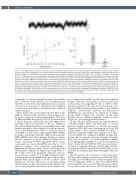
Figure 1. Electrophysiological properties of cation channels in hereditary spherocytosis red cells. (A) A representative current trace recorded at -Vp = -25mV from an on-cell patch recording from a red cell of patient HS11 with hereditary spherocytosis (HS) carrying the novel heterozygous SPTB missense variant R1255G (Polyphen-2 score 0.999). Identical bath and pipette fluid composition included (in mM) 140 NaCl, 4 KCl, 1 CaCl2, 1 MgCl2, 10 NaHEPES at a final pH of 7.40. On-cell patch currents were recorded by an Axopatch 200b amplifier and digitized by a Digidata 1440A A-D converter (Molecular Devices, Sunnyvale, CA, USA). Seal resistances were 6.0±1.0 GΩ (n=7) in non-HS cells, 5.0±0.8 GΩ (n=14) in HS cells without GsMTx4 in the pipette solution, and 4.8±1.0 GΩ (n=6) in HS cells with GsMTx4 in the pipette solution. Seal duration for recordings on HS cells unexposed to GsMTx4 was 18±11 min. Data were filtered at 500 Hz, digitized at 2 kHz by PClamp and analyzed offline by Clampfit (PCLAMP11, Molecular Devices). (B) Current-voltage relationship of HS11 red cell current meas- ured in a representative on-cell patch, with unitary slope conductance of 21 pS. The current-voltage (I-V) relationship was generated in Clampex (PCLAMP 11, Axon Instruments) with the real-time control window in gap-free mode to record current traces of 10–30 s duration. Test potentials were selected in 25-50 mV increments ranging between a minimum of -100 mV to a maximum of +100 mV. (C) Summary data for NPo calculated from on-cell patch current traces of 5- 10 s duration recorded in 16 cells from six HS mutant genotypes and in six cells from three mutant HS genotypes in the additional presence of GsMTx4 (1 μM) in the pipette fluid. NPo values recorded in seven non-HS red cells from four normal individuals (AA) are also shown. *P<0.05 for the t-test comparing normal to HS cells, and for the Mann-Whitney test comparing HS cells in the presence versus absence of GsMTx4.
centrations of fluorometrically measured intracellular [Ca2+].5 However, Petkova-Kirova et al.6 recently reported that HS red cells of the same individuals had a spectrum of decreased, increased, or unchanged cation channel activities as measured by an automated whole cell patch clamp technique.
These studies led us to investigate whether HS red cells might be characterized by increased cation channel activ- ity as detected by on-cell patch clamp analysis. We isolat- ed DNA and RNA from whole blood of 13 patients with a clinical diagnoses of HS under protocols approved by Investigational Review Boards of Boston Children’s Hospital and Beth Israel Deaconess Medical Center. The hematologic indices of the patients’ red cells are present- ed in Online Supplementary Table S1. From the isolated total RNA, we generated complementary DNA (cDNA) for Sanger sequencing of SLC4A1. cDNA and/or genomic DNA (gDNA) from those patients lacking an evident SLC4A1 mutation in blood cDNA was subjected to Sanger sequencing of ANK1 and SPTB. Whole exome sequencing was reserved for gDNA from the six of 13 patients’ samples that remained uninformative. Mutations detected by whole exome sequencing were subsequently confirmed by Sanger sequencing (Table 1). We found seven novel pathogenic mutations and one novel missense variant of very high predicted patho- genicity in previously identified HS genes among these patients with clinical diagnoses of HS. A subset of HS mutant red cells was subjected to on-cell patch clamp analysis (Figure 1). All cells in which stable gigohm seals were achieved exhibited substantial cation channel activ-
ity as compared to non-HS red cells. Mean cation channel unitary conductance among HS red cells was 26±2.1 pS (n=6 genotypes encompassing 16 cells; see Table 1). This increased activity, in the cases tested, was nearly com- pletely inhibited by the mechanosensitive cation channel inhibitor, Grammastola spatulata mechanotoxin-4 (GsMTx4) at a concentration of 1 mM in the recording pipette (Table 1, Figure 1C). Non-HS red cells from healthy donors exhibited minimal channel activity (Figure 1C), as we had previously reported.7
In this collection of HS patients, we found mutations in SLC4A1, ANK1, SPTA1, and SPTB (Table 1). Several patients exhibited mutations in SLC4A1 previously reported in HS. HS2 was heterozygous for both HS mutant Band 3 Lyon (SLC4A1 R150X) and Band 3 Montefiore (SLC4A1 E40K). Each mutation was unde- tected in cDNA but confirmed in gDNA, strongly sug- gesting that the mutant transcript carrying both muta- tions was a substrate of nonsense-mediated mRNA decay. Siblings HS3 and HS4 were each heterozygous for Band 3 Bicetre (SLC4A1 R490C). HS5 was heterozygous for Band 3 Osnabruck (SLC4A1 del663). HS6 was het- erozygous for Band 3 Prague III (SLC4A1 R870W), accom- panied by the nonpathogenic Band 3 Memphis I (SLC4A1 E56K).
Our HS patients also revealed a novel mutation in SLC4A1 and several novel mutations in ANK1 and SPTB, including a novel SPTB missense variant strongly predict- ed to be pathogenic (Table 1). The novel SLC4A1 E68X mutation in HS1 was associated with nonsense-mediated decay, whereas the known rare SLC4A1 R180H variant
2760
haematologica | 2021; 106(10)
