
Page 231 - 2021_10-Haematologica-web
P. 231
Letters to the Editor
A Grammastola spatulata mechanotoxin-4 (GsMTx4)-sensitive cation channel mediates increased cation permeability in human hereditary spherocytosis of multiple genetic etiologies
Hereditary spherocytosis (HS) is the most common inherited hemolytic anemia among people of Northern European descent. HS is caused by mutations in genes encoding the erythroid cytoskeleton proteins ankyrin-1 (ANK1), b-spectrin (SPTB), and α-spectrin (SPTA1), the major intrinsic erythroid membrane protein and chloride- bicarbonate exchanger, SLC4A1/band 3, and rarely EPB42/protein 4.2. These mutations lead to destabiliza- tion and progressive loss of red cell membrane lipids and surface area, and in some cases to destabilization of cytoskeletal-membrane attachment. The resulting red cells often exhibit normochromic or hyperchromic, mild- moderate microcytosis with increased incubated osmotic fragility and reduced deformability. Anemia and chronic hemolysis can be accompanied by hyperbilirubinemia
and painful splenic enlargement. Splenectomy often pro- vides symptomatic relief and attenuates anemia and hemolysis.1
Increased erythroid cation permeability in HS was first reported by Harris and Prankerd (1953) and Bertles (1957), as subsequently cited by Zarkowsky et al.2 Later reports of increased red cell cation permeability appeared in the setting of Southeast Asian ovalocytosis and cyro- hydrocytosis in association with SLC4A1 mutations, in the setting of overhydrated stomatocytosis in association with mutations in RHAG, SLC2A1, and SLC4A1, and in the setting of familial pseudohyperkalemia associated with ABCB6 mutations.3 Spherocytic mouse red cells genetically lacking EBP41 or EBP42, or haploinsufficient for SLC4A1 exhibited enhanced Gardos channel activity and increased hemolysis in the presence of the Gardos inhibitor, clotrimazole,4 consistent with enhanced non- specific cation permeability associated with these mouse HS models. Human HS red cells of diverse genotype were uniformly characterized by increased steady-state con-
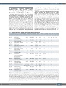
Table 1. Hereditary spherocytosis mutations accompanied by increased cation currents.
Subject Genetic diagnosis# Ref. Sanger sequence
HS1 (M) SLC4A1 E68X, novelf cDNA, gDNA
NPo NPo Unitary +GsMTx4 Conductancea
n.d. n.d. n.d.
Fam Osm Hxb Frgc
n.a n.a.
Tx/ Spx/ Acd Cxe
n.a. n.a.
HS2 (F)
HS3, HS4 (M,F; sibs) HS5 (F)
HS6 (F) HS7 (F) HS8 (F)
HS9 (M)
HS10 (F)
HS11 (F)
HS12 (M)
HS13 (M)
c.202G>T w NMD%
SLC4A1 R150X, c.448C>T,
Band 3 Lyon w NMD%
SLC4A1 R490C, c.1648C>T,
Band 3 Bicetre
SLC4A1 M663del, c.1987-9del*,
Band 3 Osnabruck II
SLC4A1 R870W, c.2608C>T,
Band 3 Prague III
ANK1 E883Gfs32X,
c.2648delA, w NMD%
(g) (h) (i) (j) novelk
35 pS 28 pS 25 pS n.d. n.d. 22.5 pS
25.8 pS
n.d.
21 pS
n.d.
n.d.
cDNA, gDNA cDNA gDNA cDNA cDNA, gDNA cDNA, gDNA
cDNA, gDNA cDNA, gDNA gDNA
novel cDNA, gDNA novel cDNA, gDNA
1.43 0.14 (n=2) (n=1) 3.54 n.d.
(n=1) 0.91±0.32 n.d.
(n=4)
n.d. n.d.
n.d. n.d.
0.56±0.13 0.059 (n=4) (n=1)
0.93±0.17 n.d. (n=4)
n.d n.d.
1.26 0.042±0.02 (n=1) (n=4)
n.d. n.d. n.d. n.d.
+ n.a - + + +- + + + + - + +/– – – + n.a. + + + + + +
++ - -
++ + +
+ n.a. - -
++ + -
- + + +
A1110-Q1111del, novel
c.3328-3333del6, w pNMD%%
(Exon 28 mutant alters splice acceptor site)
ANK1 K1140Gfs86X, c.3416ins16, w pNMD%% ANK1 E1289Gfs86X, c.3865dupG, w NMD% SPTB R1255G, c.3763A>G
SPTB G1450Rfs40X,
c.4346dupG, w NMD%
SPTB E1815Pfs90X,
c.5443G>CC, w NMD%
novell
novel
novel variant, likely pathogenicm
#cDNA numbering from initiator ATG of the open reading frame; SLC4A1: NP000333.1, NM000342.4; ANK1 isoform 1: NP065209, NM020476.2; SPTA1: NP003117, NM003126.4; SPTB erythrocyte isoform A: NP 001020029.1; var. 1 NM001024858. %complete nonsense-mediated decay (NMD); %%partial nonsense-mediated decay (pNMD); *deletion of any three consecutive nucleotides between c.1987-1992; n.d.: not done; n.a.: not available. aUnitary slope conductance measured in a single representative cell of specified genotype, without GsMTx4 in the pipette solution. bFamHx: family history of hereditary spherocytosis; cOsmFrg: results of osmotic fragility test; dTx/AC: history of transfusion without or with aplastic crisis; eSpx/Cx: history of splenectomy or cholecystectomy. fFound with SLC4A1 R180H, c.539G>A, rs147390654, MAF 0.0001-0.01 in different popu- lations, detected in cDNA and gDNA. gAlloisio N et al. Blood 1996;88:1062-1069, conallelic with SLC4A1 E40K, c.118G>A, Band 3 Montefiore. Rybicki AC et al. Blood 1993;81:2155-2165.Detected in cDNA and gDNA.hDhermy D et al. Br J Haematol 1997;98:32-40.iEber SW et al.Nat Genet 1996;13:214-218. jJarolim P et al.Blood 1995;85:634- 640, found together with benign variant SLC4A1 K56E, c.166A>G, Band 3 Memphis.Yannoukakos D et al. Blood 1991;78:1117-1120. Detected in cDNA and gDNA. kFound together with SPTA1 R1074H,c.3221G>A,rs551084590,MAF 0.00004,detected in gDNA.lFound together with SLC4A1 P854L,c.2561C>T;and K56E,c.166A>G,Band 3 Memphis II, Bruce LJ et al. J Biol Chem 1994;269:16155-16158. Detected in cDNA. mFound together with likely benign variant ANK1 R619H, c.1856G>A, rs2304877, Ankyrin Bruggen, Nakanishi H et al. Int J Hematol 2001;73:54-63. Detected in gDNA.
haematologica | 2021; 106(10)
2759
