
Page 160 - 2021_10-Haematologica-web
P. 160
J.E. Ramis-Zaldivar et al.
D
E
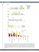
Figure 2. Genetic alterations of plasmablastic lymphoma. (A) Global copy number and (B) copy number neutral loss of heterozygosity (CNN-LOH) profile of 33 cases of plasmablastic lymphoma (PBL). The x-axis indicates chromosomes from 1 to Y and p to q. The y-axis indicates the frequency of the genomic aberration among the cases analyzed. Gains are depicted in blue, losses are depicted in red and CNN-LOH are depicted in yellow. Recurrent copy number and CNN-LOH regions (>20% of cases) are indicated. (C) Bar plot showing genes mutated in more than 5% of 27 cases of PBL. The color of the bar indicates the Epstein-Barr virus (EBV) infection status. (D) A diagram of the relative positions of driver mutations is shown for STAT3, TP53, NRAS and MYC genes. The x-axes indicate amino acid positions. CCD: coiled coil domain; DBD: DNA-binding domain; TAD: transcription activation domain; HLH: helix-loop-helix; LZ: leucine zipper. (E) Recurrent mutated pathways in 27 PBL. The bar graph shows the total number of mutated cases for each pathway. Asterisks represent significant mutated pathways in EBV-negative (n=9) versus pos- itive (n=14) PBL.
2688
haematologica | 2021; 106(10)
