
Page 149 - 2021_10-Haematologica-web
P. 149
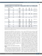
Evolution of ISFN to aggressive B-cell lymphoma
Table 2. Cell of origin, BCL2 translocation and immunoglobulin gene analysis.
Case Diagnosis Cell of origin based BCL2 on Hans algorithm FISH
De novo aggressive B-cell lymphoma
BCL2 breakpoint
MBR-JH MBR-JH
Neg Neg
MBR-JH† MBR-JH MBR-JH
Neg Neg
Neg Neg
MBR-JH MBR-JH
MBR-JH MBR-JH MBR-JH
3’MBR-JH 3’MBR-JH
MBR-JH† MBR-JH MBR-JH
Clonality*
Mono Mono
Mono Mono
Poly Poly Poly
Mono Mono
Poly Poly
Poly Mono
Mono Mono Mono
Mono Mono
Mono Mono Mono
IGH V/J usage
V3/J4 V3/J4
V2/J4 V2/J4
— — —
V3/J6 V3/J6
— —
— V3/J3
V3/J6 V3/J6 V3/J6
V3/J3 V3/J3
V3/J4 V3/J4 V3/J4
V1/J6 V1/J6 V1/J6
Glycosylation site Location Motif#
Status
Clonally related Clonally related Clonally related
Clonally related Clonally related§ Clonally related
Clonally related
Clonally related Clonally related
Clonally related
1 ISFN HGBL-TH
2 ISFN DLBCL
3 ISFN DLBCL
DLBCL
4 ISFN DLBCL
5 ISFN DLBCL
6 ISFN DLBCL
Transformed FL
7 ISFN FL
DLBCL
8 ISFN HGBL-DH
9 ISFN FL
HGBL-DH
10 ISFN FL
DLBCL
— + GCB +
— + GCB +
— + GCB + GCB +
— + GCB +
— +f GCB +f
— + GCB +
— +
— + GCB +
— + GCB +
— +
— + GCB +
— +
— + GCB +
CDR3 CDR3
CDR3 CDR3
CDR3 CDR3
FR3
CDR3 CDR3 CDR3
— — —
— —
—
NLS NLS
NDS NTS
NAS NAS
NLT
NLT NLT NLT
MBR-JH Mono“ MBR-JH Mono“ MBR-JH Mono“
FR2/CDR2 NIT None (FR1 PCR)
CDR3 NCS CDR3 NCS None (FR2 PCR)
CDR3 NFS CDR3 NYS CDR3 NYS
CDR: complementarity-determining region; FR: framework region; Neg: negative (i.e., no BCL2 rearrangement detected by polymerase chain reaction [PCR]);V/J: variable/join- ing gene segment; ISFN: in situ follicular neoplasia ; FL: follicular lymphoma; HGBL-TH: high-grade B-cell lymphoma triple-hit; HGBL-DH: HGBL double-hit; DLBCL: diffuse large B-cell lymphoma; LN: lymph node; MBR: major breakpoint region; GCB: germinal center B-cell-like; IGH: immunoglobulin H. *Based on Lymphotrack and/or GeneScan analysis. #Single-letter amino acid code.†Amplified with breakpoint-specific primers.fDemonstrated using an IGH/BCL2 dual-color,double fusion probe.§Based on the demonstration of shared mutations.“Demonstrated with clone-specific primers.
sites in seven of seven ISFN, three of three FL and six of eight aggressive BCL. In three cases (cases 1, 4, and 7), the ISFN and their transformed counterpart(s) demonstrated identical glycosylation sites, whereas two ISFN (cases 2 and 10) showed motifs at the same location, but with a different sequence than those exhibited by the clonally related manifest lymphomas. Moreover, two HGBL lacked N-glycosylation sites, although novel motifs were detected in the related ISFN (cases 8 and 9) and FL (case 9) lesions. Intraclonal heterogeneity of the clonal IGH re- arrangement was present in all types of samples. However, heterogeneity was more pronounced in the precursor lesions, as evidenced by more evenly distrib- uted subclones, whereas DLBCL and HGBL samples exhibited one or two subclones that were highly domi- nant. Phylogenetic trees constructed for five cases demonstrated separate clustering of ISFN and DLBCL/HGBL sequences indicative of divergent evolu- tion (Online Supplementary Figure S1).
Mutational analysis reveals distinct clonal evolution patterns
BCL2 was the most frequently mutated gene, with all samples harboring at least one non-synonymous, synony- mous or 5’UTR mutation, although most samples, includ- ing seven of ten ISFN lesions, demonstrated several BCL2 mutations (Figure 2; Online Supplementary Tables S4 and S5). Other recurrently mutated genes were CREBBP (11 of 24 samples), KMT2D (11 samples), and EZH2 (ten sam-
ples), as well as TNFRSF14, IGLL5, and GNA13. In ISFN lesions, mutations in chromatin modifying genes remained the most frequent alterations, with five samples showing a CREBBP mutation. In contrast, TP53 (four samples), CD79B (one sample) and HIST1H1B (one sam- ple) were exclusively altered in the aggressive compo- nents. All TP53 mutations were located in the DNA bind- ing domain, with variant allele frequencies ranging from 52% to 84%, indicating a loss of the second allele. For two samples (cases 1 and 2), this was confirmed by FISH.
Of the nine cases clonally related by IG and/or BCL2 breakpoint sequence analysis, all but one (case 1) demon- strated shared mutations between the ISFN and the trans- formed counterpart(s), ranging from one to six shared mutations per paired samples. For example, the DLBCL of case 4 had four non-synonymous mutations of BCL2, TNFRSF14, HIST1H1D and EP300 in common with the ISFN that was present 159 months prior. However, ISFN and DLBCL of case 5 also exhibited matching KMT2D p.(Q4473*) and IGLL5 p.(C3S) mutations, demonstrating their clonal relationship despite the lack of a detectable clonal IG re-arrangement or BCL2 translocation sequence. All investigated FL showed more than one mutation shared with both the ISFN and the aggressive BCL. Mutations only present in the clinically manifest lym- phomas were observed in all cases with the exception of one FL (case 9). Nevertheless, six ISFN lesions also carried private variants that were not identified in their clonally related counterparts, indicating early divergence. The
haematologica | 2021; 106(10)
2677
