
Page 120 - 2021_10-Haematologica-web
P. 120
G. Zuchtriegel et al.
Discussion
The trafficking of circulating leukocytes from the venular microvasculature to the site of injury or infection is a key event in the pathogenesis of inflammatory diseases.11-14 The role of the matricellular protein VN in this fundamental biological process is still unclear. Under homeostatic con- ditions, VN predominantly circulates in the blood as a monomer. In the acute inflammatory response, however, the binding of activated PAI-1 to VN monomers induces conformational changes in this macromolecule that effec- tively promote its multimerization. This allows multi- meric VN to specifically bind to the surface of endothelial cells and, in turn, to unfold previously cryptic binding sites as evidenced by different in vitro studies.30-32 Accordingly, we found VN to be deposited on the luminal surface of the venular microvasculature in inflamed tis- sue. Since GAG cover the luminal aspect of microvascular
endothelial cells and have previously been reported to interact with VN through its core polypeptide,33 we hypothesized that these carboanhydrates serve as bind- ing partner for VN on the inner vessel wall. Confirming this assumption, enzymatic degradation of GAG almost completely abrogated the deposition of VN on the acti- vated microvascular endothelium, thus suggesting that VN is immobilized in inflamed tissue on the surface of microvascular endothelial cells by endothelial GAG.
In order to evaluate the functional relevance of VN for leukocyte migration to the site of inflammation, we employed a peritoneal leukocyte trafficking assay. In these experiments, neutrophil extravasation to the peri- toneal cavity was severely compromised in VN-/- mice as compared to WT controls. Notably, this impairment in neutrophil recruitment reached similar levels in heterozy- gous and homozygous VN-/- mice suggesting that compar- atively large amounts of endothelially deposited VN are
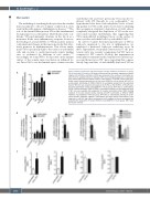
Figure 5. Effect of vitronectin and plasminogen activator inhibitor-1 heteromers on activa- A tion of neutrophil b2 integrins. (A) Using multi-channel flow cytometry, expression of the b2 integrins LFA-1/CD11a and Mac-1/CD11b was analyzed on the surface of neutrophils iso- lated from the peripheral blood of wild-type (WT) mice undergoing exposure to vitronectin (VN), plasminogen activator inhibitor-1 (PAI-1), uPA, VN-PAI-1, or VN-uPA, panels show quan- titative results (mean±standard error of the mean [SEM] for n=4 per group; #P<0.05 vs. unstimulated). (B) Binding of ICAM-1/CD54-Fc to neutrophils isolated from the peripheral blood of WT mice was analyzed upon exposure to VN, PAI-1, uPA, VN-PAI-1, or VN-uPA, pan- els show quantitative results (mean±SEM for n=6 per group; #P<0.05 vs. unstimulated). VN-PAI-1-elicited binding of ICAM-1/CD54 to neutrophils isolated from the peripheral blood of WT mice was analyzed after application of receptor associated protein (RAP; blocking receptors of the LDL receptor family), blocking anti-LRP-1 antibodies, or different MAPK inhibitors (mean±SEM for n=4-6 per group; #P<0.05 vs. unstimulated). Binding of confor- mation-specific antibodies ‚KIM127‘ (intermediate and high affinity conformations of b2 integrins) or ‚mAB 24‘ (high affinity conformation of b2 integrins) to human neutrophils was analyzed after application of human VN-PAI-1 (mean±SEM for n=3 per group). MFI: mean
fluorescent intensity.
B
C
2648
haematologica | 2021; 106(10)
