
Page 247 - 2021_09-Haematologica-web
P. 247
Case Report
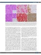
Figure 1. Representative pathologic findings of sequential grey zone lymphomas (Case #1). Initial cervical lymph node biopsy shows classic Hodgkin lymphoma (upper panel, A to C). (A) Characteristic Hodgkin and Reed–Sternberg (HRS) cells are present in a polymorphous inflammatory background (hematoxylin and eosin stain[H&E]); the HRS cells are negative for EBER (inset A). (B) The neoplastic HRS cells are positive for CD30 (red, membranous) and weekly positive for PAX-5 (brown, nuclear). (C) They are negative for CD20. (D to F) Lesional brain biopsy shows sequential central nevous system large B-cell lymphoma (lower panel). (D) Diffuse sheets of large lymphoma cells shows centrally located prominent nucleoli (H&E); they are negative for EBER (inset D). (E) The lymphoma cells are diffusely positive for CD30 (red, membranous) with strong nuclear PAX-5 (brown) expression, (F) and express strong and homogeneous CD20.
ation sequencing (NGS) using a capture-based 152 gene custom-designed hematologic malignancy panel was per- formed on paired cHL and CNS LBCL tumors to assess for genomic alterations as previously described.5 These stud- ies were performed under institutional-approved study protocols. We present the clinicopathologic and genomic features of the paired lesions in this previously unreported presentation of pediatric sequential GZL.
Case #1, 17M. A 17 year-old male presented with large mediastinal and supraclavicular masses with disseminat- ed spleen, liver, and bone lesions. Left cervical lymph node sampling revealed the classic histology and immunophenotype of NS-cHL (Figure 1A to C, Table 1). A concomitant outside bone marrow sample performed approximately 3 weeks prior revealed LBCL with sizable clusters of large lymphoma cells, consistent with a diag- nosis of synchronous GZL. Staging bone marrow was negative for involvement by lymphoma. Complete remis- sion was achieved after initial treatment with ABVE-PC chemotherapy regimen. Six months, after initial diagnosis (2 months post-therapy), several supra- and infra-tentorial brain lesions and extensive leptomeningeal disease appeared. A biopsy of a CNS lesion revealed diffuse sheets of large lymphoma cells having open chromatin, prominent centrally located nucleoli, and a moderate amount of clear to eosinophilic cytoplasm. The lym- phoma cells showed diffuse and strong expression for CD45, CD20, PAX-5, CD30, and expression of CD79a, and were negative for CD15, EBER, and ALK (Figure 1D to F, Table 1). A diagnosis of BCL-U-IND consistent with sequential GZL was rendered. He was treated according to POG9917 Arm A as a bridge to bone marrow trans- plant with a mismatched unrelated donor and received total body irradiation (450 cGy). He was alive at 81.7 months follow-up.
Case #2, 16F. A 16 year-old female presented with a large mediastinal mass with cervical lymphadenopathy and multiple bilateral renal and pulmonary nodules. NS- cHL was diagnosed from cervical lymph node biopsy; staging bone marrow was negative. She achieved com- plete remission after ABVE-PC and radiotherapy to the mediastinal mass and other slow-responding areas of dis- ease. Seven months after initial diagnosis (2 months post- therapy), a solitary right temporal lesion was identified. A biopsy revealed essentially similar morphologic and immunophenotypic findings to the CNS lesion of case #1 (Table 1), and a diagnosis of BCL-U-IND, consistent with sequential GZL was rendered. She was treated with ANHL1131 Group C1 and surgical excision and was alive at 13.5 months follow-up.
Molecular findings. The microarray and NGS results are summarized in Table 2. In both NS-cHL, near-diploid male or female genomes and no variants of established or potential clinical significance (Tier I/II, Table 2) were detected consistent with “negative” genomic profiles reported in bulk cHL lesions without Reed-Sternberg cell enrichment.6,7 In case #2, a shared 3.0 MB region of copy- neutral loss of heterozygosity (LOH) in chromosome 1p36.11-p35.3 was observed that was most likely germline in origin. Both CNS LBCL harbored complex cytogenomic arrays including 2p16.1 and 9p24.1 gains (detected in both cases, Table 2, denoted by *) and 16p13.3 copy-number abnormalities (case #2 only). LOH of chromosome 6p and gain of chromosome 12p were also observed in both CNS LBCL (Table 2, denoted by *). NGS revealed shared NS-cHL/CNS LBCL variants of uncertain significance (VUS, Tier III) in CREBBP p.T974N (case #1) and RELN p.N352S and KMT2D p.E4694Q (case #2). The sequential CNS LBCL in case #1 harbored addi-
haematologica | 2021; 106(9)
2535
